β-Actin Rabbit Monoclonal Antibody
| Cat Number: | MAB026 |
|---|---|
| Conjugate: | Unconjugated |
| Size: | 300 ug |
| Clone: | 13E5 |
| Concentration: | 1mg/ml |
| Host: | Rabbit |
| Isotype: | IgG |
| Immunogen: | Recombinant protein.This information is considered to be commercially sensitive. |
| Reactivity: | Human,Mouse,Rat,Chicken,Zebrafish,Pig,Cow |
| Applications: | Western Blot: 1:10000-1:100000 Immunofluorescence: 1:200-1:800 Immunocytochemistry: 1:200-1:800 Immunohistocehmistry(paraffin-embedded tissues): 1:3000 - 1:10000 ELISA |
| Molecular: | 42kDa |
| Purification: | Affinity purification |
| Synonyms: | BRWS1; PS1TP5BP1; β-Actin |
| Background: | This gene encodes one of six different actin proteins. Actins are highly conserved proteins that are involved in cell motility, structure, integrity, and intercellular signaling. The encoded protein is a major constituent of the contractile apparatus and one of the two nonmuscle cytoskeletal actins that are ubiquitously expressed. Mutations in this gene cause Baraitser-Winter syndrome 1, which is characterized by intellectual disability with a distinctive facial appearance in human patients. Numerous pseudogenes of this gene have been identified throughout the human genome. Note:Due to the high antibody titer, it is advisable to be diluted before use. Please dilute 5μL of the antibody solution with 45μL of PBS solution, containing 50% glycerol. The diluted antibody can be stored at -20°C. |
| Form: | liquid |
| Buffer: | PBS containing 50% glycerol and 0.05% BSA, preserved with proclin300 or sodium azide (as specified on the Certificate of Analysis), pH 7.3. |
| Storage: | Store at -20℃. Avoid freeze / thaw cycles. |
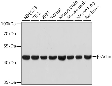
Western blot analysis of various lysates using β-Actin Rabbit mAb at 1:100000 dilution.
Secondary antibody: HRP-conjugated Goat anti-Rabbit IgG (H+L) at 1:10000 dilution.
Lysates/proteins: 25μg per lane.
Blocking buffer: 3% nonfat dry milk in TBST.
Detection: ECL West Pico Plus.
Exposure time: 30s.
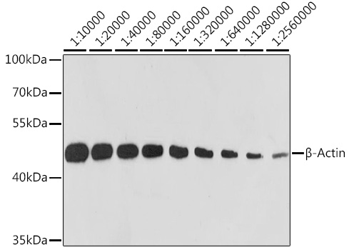
Western blot analysis of lysates from HeLa cells, using β-Actin Rabbit mAb at 1:10000-1:75000 dilution.
Secondary antibody: HRP-conjugated Goat anti-Rabbit IgG (H+L) at 1:10000 dilution.
Lysates/proteins: 25μg per lane.
Blocking buffer: 3% nonfat dry milk in TBST.
Detection: ECL West Pico Plus.
Exposure time: 10s.
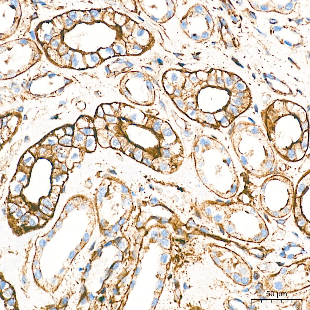
Immunohistochemistry analysis of paraffin-embedded Human kidney tissue using β-Actin Rabbit mAb at a dilution of 1:10000 (40x lens). High pressure antigen retrieval performed with 0.01M Tris-EDTA Buffer (pH 9.0) prior to IHC staining.