
Anti-Iba1 Mouse Monoclonal Antibody
| Cat Number: | MABS12105 |
|---|---|
| Conjugate: | Unconjugated |
| Size: | 100 ug |
| Clone: | 5G4E7 |
| Host: | Mouse |
| Isotype: | IgG1, κ |
| Immunogen: | Recombinant protein corresponding to Mouse Iba1 |
| Reactivity: | Human,Mouse,Rat,Monkey |
| Applications: | IHC(PFA fixed)/IF(PFA fixed) 1: 500-1: 4000 Species: Human, Mouse, Rat, Monkey Positive tissue: brain |
| Purification: | Affinity purification |
| Synonyms: | AIF-1, IBA, IRT-1, IRT1, allograft, AIF-1, AIF1, G1, IBA1, IRT 1, Protein G1, Ionized calcium-binding adapter molecule 1, MRF1, Microglia response factor, BART 1, AIF1 protein |
| Background: | AIF1, also named as G1, IBA1 or daintain/AIF-1, is a 143 amino acid cytoplasmic, inflammation response scaffold protein. It is constitutively expressed in monocytes and macrophages and is known to be involved in macrophage activation. It is a marker of activated macrophage. Despite a lack of detailed knowledge on the in vivo physiological functions of AIF-1, there is growing evidence that shows its aberrant expression contributes to the pathogenesis of many autoimmune diseases, including rheumatoid arthritis. AIF1 is an actin-binding protein that enhances membrane ruffling and RAC activation. It enhances lymphocyte migration and the actin-bundling activity of LCP1. |
| Buffer: | PBS with 0.15% ProClin300, 100 μg/mL BSA and 50% glycerol |
| Storage: | Store at -20℃ for one year. Avoid repeated freeze/thaw cycles. |
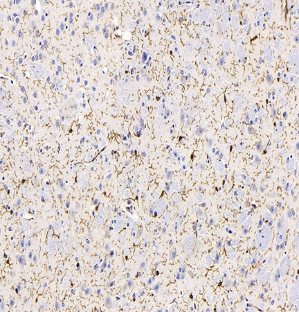
IHC analysis of Iba1. Sample: Mouse brain, 4% PFA 12-24h. Antigen retrieval: Citrate buffer (pH 6.0), 98℃, 20 min. Primary antibody: 1: 2000, 4℃ overnight. Secondary antibody: HRP conjugated Goat Anti-Mouse lgG (H+L) 1: 200, RT, 1h.
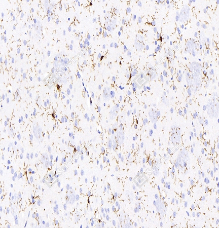
IHC analysis of Iba1. Sample: Rat brain, 4% PFA 12-24h. Antigen retrieval: Citrate buffer (pH 6.0) 98℃, 20 min. Primary antibody: 1: 2000, 4℃ overnight. Secondary antibody: HRP conjugated Goat Anti-Mouse lgG (H+L) 1: 200, RT, 1h.
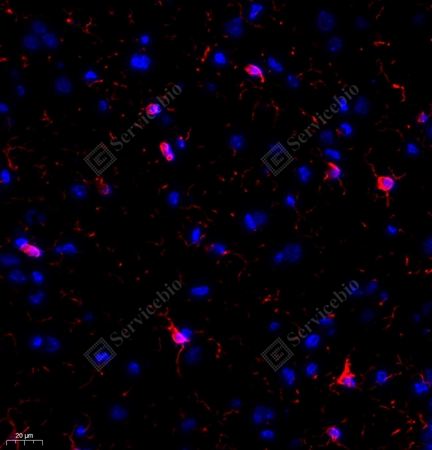
IF analysis ofIba1 red. Sample: Rat brain, 4% PFA12-24h. Antigen retrieval: Tris-EDTA buffer (pH 9.0), 98℃, 20min. Blocking buffer: 3% BSA in PBS, RT, 30min. Primary antibody: 1: 1000, 4℃ overnight. Secondary antibody: Cy3 conjugated Goat Anti-Mouse IgG (H+L), 1: 300, RT, 1h.