
Arrestin-1 Mouse Monoclonal Antibody
| Cat Number: | MAB-94385 |
|---|---|
| Conjugate: | Unconjugated |
| Size: | 100 ug |
| Clone: | S128 |
| Concentration: | 1mg/ml. |
| Host: | Mouse |
| Isotype: | IgG1 |
| Immunogen: | Recombinant bovine arrestin 1 with the first 20 amino acids of the C-terminus truncated |
| Reactivity: | Human, rat, mouse, cow, pig, horse |
| Applications: | WB: 1:5,000. IF/ICC: 1:1,000. IHC: 1:1,000. |
| Molecular: | 48kDa |
| Purification: | Affinity Purified |
| Synonyms: | ARRB1; ARR1; Beta-arrestin-1; Arrestin beta-1 |
| Background: | The MCA-S128 antibody was made against recombinant bovine arrestin 1 with 20 N-terminal amino acids truncated (7). The antibody works well for western blotting and for IF, ICC and IHC (for IHC see data under “Additional Data” tab). The antibody does not cross-react with cone-arrestin, βarrestin 1 or β-arrestin 2. The MCA-S128 antibody can be used to study this protein in cells, tissues and tissue lysates. |
| Form: | Liquid |
| Buffer: | Protein G affinity purified antibody at 1mg/mL in 50% PBS, 50% glycerol plus 5mM NaN3 |
| Storage: | Store at 4°C for short term, for longer term store at -20°C. Stable for 12 months from date of receipt. |
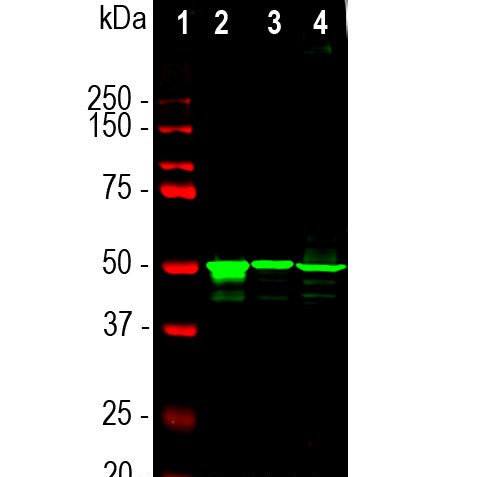
Western blot analysis of retina lysates from different species using mouse mAb to arrestin 1, MCA-S128, dilution 1:5,000 in green: [1] protein standard (red), [2] rat, [3] mouse, and [4] cow retina lysates. The Arrestin 1 antibody detects arrestin 1 a.k.a. visual arrestin and Santigen, running at about 48kDa.
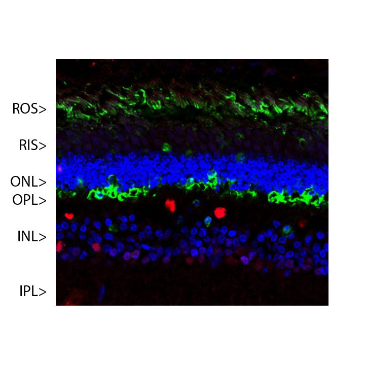
Immunofluorescent analysis of a pig retina section stained with mouse mAb to arrestin 1, dilution 1:1,000, in green, and costained with chicken pAb to FOX2, CPCAFOX2 dilution 1:1,000 in red. The blue is Hoechst staining of nuclear DNA. Arrestin 1 antibody reveals visual arrestin1 protein in cells located in outer segments of photoreceptors layer (OS) and in outer plexiform layer
(OPL) of retina. FOX2 antibody stains nuclei of horizontal and other neurons in the INL and IPL