BDNF
| Cat Number: | AB-82598 |
|---|---|
| Conjugate: | Unconjugated |
| Size: | 100 ug |
| Clone: | POLY |
| Concentration: | 1mg/ml |
| Host: | Rb |
| Isotype: | IgG |
| Immunogen: | E.coli-derived human BDNF recombinant protein (Position: H129-R247). Human BDNF shares 100% amino acid (aa) sequence identity with both mouse and rat BDNF. |
| Reactivity: | Hu,Ms,Rt |
| Applications: | • Western Blot : 1:500 – 1:1000 |
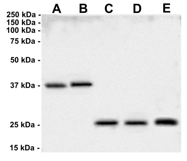
| Molecular Weight: | 37 kDa , 25 kDa |
| Purification: | Aff. Pur. |
| Background: | During development, promotes the survival and differentiation of selected neuronal populations of the peripheral and central nervous systems. Participates in axonal growth, pathfinding and in the modulation of dendritic growth and morphology. Major regulator of synaptic transmission and plasticity at adult synapses in many regions of the CNS. The versatility of BDNF is emphasized by its contribution to a range of adaptive neuronal responses including long-term potentiation (LTP), long-term depression (LTD), certain forms of short-term synaptic plasticity, as well as homeostatic regulation of intrinsic neuronal excitability. |
| Form: | Liquid |
| Buffer: | Each vial contains 5mg BSA, 0.9mg NaCl, 0.2mg Na2HPO4, 0.05mg NaN3. |
| Storage: | 4° C for 2 months for longer terms at -20°C. Avoid Freeze and thaw cycles. |

Prof.Rosa Di Liddo , Dr. Thomas Bertalot, Dr. Sandra Schrenk
University of Padova
Department of Pharmaceutical and Pharmacological Sciences
See western blot protocol on Western Blotting AB-82598
(Protocol of Prof.Rosa Di Liddo,Dr.Thomas Bertalot, Dr. Sandra Schrenk)
University of Padova
Department of Pharmaceutical and Pharmacological Sciences
Sample preparation
Whole protein lysate was obtained using RIPA lysis buffer supplemented with protease inhibitors. All procedures were performed on ice and homogenization of samples was executed using a 22G needle syringe.
1. SDS-PAGE Electrophoresis: Sample loaded: 10μg – Poly-Acrylamide gel at 12%
2. Transfer: 0.45μm PVDF Membrane (Transfer for 80 min at 4°C on ice) -140 Volts and 14 mA
3. Blocking: – Blocking solution: 5% no-fat milk with TBS -Time: 2h
4. Incubation of Primary antibody: Dilution of primary antibody: 1:1000 [1μg/mL] -Dilute buffer: 1% no-fat milk with
TBS – Time and temperature: 4°C overnight in constant slow shacking
5. Washing: Wash with TBS-T (Tween-20, 0.25%) for three times (15 min, 5 min, 5 min)
6. Incubation of Secondary antibody: Dilution of secondary antibody: HRP-Goat anti-rabbit IgG (1:2500)
7. Dilute buffer: 1% no-fat milk with TBS -Time and temperature: RT, 90min on shaker
8. Washing: Wash with TBS-T (Tween-20, 0.25%) for four times (20 min, 5 min, 5 min, 5 min)
9. ECL chemiluminescent method:

Immunoflurescence AB-82598 (Protocol of Prof.Rosa Di Liddo,Dr. Thomas Bertalot,Dr. Sandra Schrenk)
University of Padova
Department of Pharmaceutical and Pharmacological Sciences
Procedures
1.Cell culture
ENSc were seeded on glass coverslips and maintained in standard medium for 7 days
2. Fixation
ENSc were fixed using a paraformaldehyde solution kit for 20 min at 4°C
3. Permeabilization
Samples were permeabilized using a 0.5% Triton X-100 solution prepared in PBS 1X for 15 min at RT
4.Blocking
Blocking solution: 5% BSA in PBS 1X for 45 min at RT
5. Incubation of Primary antibody
Dilution of primary antibody: 1:50 [20μg/mL]
Dilute buffer: 0.5% BSA in PBS 1X
Time and temperature: 4°C overnight
6. Washing
Wash with PBS 1X for three times
7. Incubation of Secondary antibody
Dilution of secondary antibody: PE-Goat anti-rabbit IgG (1:100) [10μg/mL]- Dilute buffer: 0.5% BSA in PBS 1X
Time and temperature: RT, 60min
8. Washing
Wash with PBS 1X for three times
9.Mounting
Mount the sample with Fluoromont G with DAPI

IHC(P): Mouse Brain Tissue
By Heat: Boiling the paraffin sections in 10mM citrate buffer, pH6.0, for 20mins is required for the staining of formalin/paraffin sections
