BMP2 Rabbit Monoclonal Antibody
| Cat Number: | MAB27101 |
|---|---|
| Conjugate: | Unconjugated |
| Size: | 100 ug |
| Concentration: | 1mg/ml |
| Host: | Rabbit |
| Isotype: | IgG |
| Immunogen: | Recombinant protein.This information is considered to be commercially sensitive. |
| Reactivity: | Human,Mouse,Rat |
| Applications: | WB 1:1000 - 1:6000 IF/ICC 1:100 - 1:800 IP 0.5μg-4μg antibody for 300μg-500μg extracts of whole cells ELISA Recommended starting concentration is 1 μg/mL. |
| Molecular: | 12-45kDa |
| Purification: | Affinity purification |
| Synonyms: | BDA2; BMP2A; SSFSC; SSFSC1; BMP2 |
| Background: | This gene encodes a secreted ligand of the TGF-beta (transforming growth factor-beta) superfamily of proteins. Ligands of this family bind various TGF-beta receptors leading to recruitment and activation of SMAD family transcription factors that regulate gene expression. The encoded preproprotein is proteolytically processed to generate each subunit of the disulfide-linked homodimer, which plays a role in bone and cartilage development. Duplication of a regulatory region downstream of this gene causes a form of brachydactyly characterized by a malformed index finger and second toe in human patients. |
| Form: | liquid |
| Buffer: | PBS with 0.09% Sodium azide,0.05% BSA,50% glycerol,pH7.3. |
| Storage: | Store at -20℃. Avoid freeze / thaw cycles. |
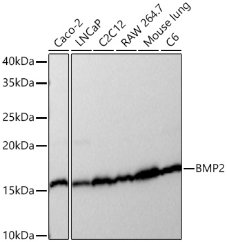
Western blot analysis of various lysates using BMP2 Rabbit mAb at 1:1000 dilution incubated at room temperature for 1.5 hours.
Secondary antibody: HRP-conjugated Goat anti-Rabbit IgG (H+L) at 1:10000 dilution.
Lysates/proteins: 25 μg per lane.
Blocking buffer: 3% nonfat dry milk in TBST.
Detection: ECL Basic Kit (RM00020).
Exposure time: 1s.
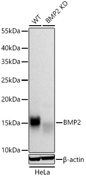
Western blot analysis of various lysates using BMP2 Rabbit mAb at 1:1000 dilution incubated overnight at 4℃.
Secondary antibody: HRP-conjugated Goat anti-Rabbit IgG (H+L) at 1:10000 dilution.
Lysates/proteins: 25 μg per lane.
Blocking buffer: 3% nonfat dry milk in TBST.
Detection: ECL West Pico Plus.
Negative control (NC): LNCaP
Exposure time: 1s.
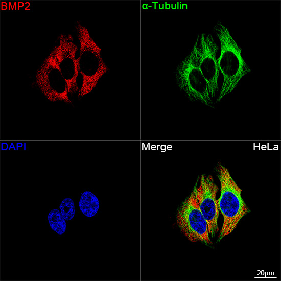
Confocal imaging of HeLa cells using BMP2 Rabbit mAb (dilution 1:100) followed by a further incubation with Cy3 Goat Anti-Rabbit IgG (H+L) (, dilution 1:500) (Red). The cells were counterstained with α-Tubulin Mouse mAb (dilution 1:400) followed by incubation with AF488-conjugated Goat Anti-Mouse IgG (H+L) Ab ( dilution 1:500) (Green). DAPI was used for nuclear staining (Blue). Objective: 100x.