CCR5 Rabbit Polyclonal Antibody
| Cat Number: | AB20261 |
|---|---|
| Conjugate: | Unconjugated |
| Size: | 100 ug |
| Concentration: | 1mg/ml |
| Host: | Rabbit |
| Isotype: | IgG |
| Immunogen: | Synthetic peptide. This information is considered to be commercially sensitive. |
| Reactivity: | Human,Mouse,Rat |
| Applications: | WB 1:500 - 1:1000 IF/ICC 1:50 - 1:200 ELISA Recommended starting concentration is 1 μg/mL. Please optimize the concentration based on your specific assay requirements. |
| Molecular: | 41kDa |
| Purification: | Affinity purification |
| Synonyms: | CKR5; CCR-5; CD195; CKR-5; CCCKR5; CMKBR5; IDDM22; CC-CKR-5; CCR5 |
| Background: | This gene encodes a member of the beta chemokine receptor family, which is predicted to be a seven transmembrane protein similar to G protein-coupled receptors. This protein is expressed by T cells and macrophages, and is known to be an important co-receptor for macrophage-tropic virus, including HIV, to enter host cells. Defective alleles of this gene have been associated with the HIV infection resistance. The ligands of this receptor include monocyte chemoattractant protein 2 (MCP-2), macrophage inflammatory protein 1 alpha (MIP-1 alpha), macrophage inflammatory protein 1 beta (MIP-1 beta) and regulated on activation normal T expressed and secreted protein (RANTES). Expression of this gene was also detected in a promyeloblastic cell line, suggesting that this protein may play a role in granulocyte lineage proliferation and differentiation. This gene is located at the chemokine receptor gene cluster region. An allelic polymorphism in this gene results in both functional and non-functional alleles; the reference genome represents the functional allele. Two transcript variants encoding the same protein have been found for this gene. |
| Form: | liquid |
| Buffer: | PBS with 0.05% proclin300,50% glycerol,pH7.3. |
| Storage: | Store at -20℃. Avoid freeze / thaw cycles. |

Western blot analysis of lysates from Rat liver, using CCR5 Rabbit pAb at 1:1000 dilution.
Secondary antibody: HRP-conjugated Goat anti-Rabbit IgG (H+L) at 1:10000 dilution.
Lysates/proteins: 25μg per lane.
Blocking buffer: 3% nonfat dry milk in TBST.
Detection: ECL Basic Kit.
Exposure time: 10s.

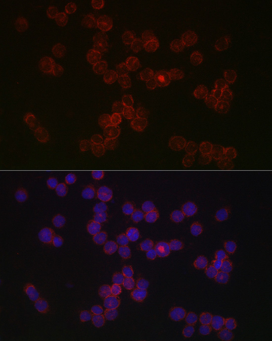
Immunofluorescence analysis of THP-1 cells using CCR5 Rabbit pAb at dilution of 1:50 (40x lens). Secondary antibody: Cy3-conjugated Goat anti-Rabbit IgG (H+L) at 1:500 dilution. Blue: DAPI for nuclear staining.

Immunofluorescence analysis of paraffin-embedded mouse spleen using CCR5 Rabbit pAb at dilution of 1:100 (40x lens). Secondary antibody: Cy3-conjugated Goat anti-Rabbit IgG (H+L) at 1:500 dilution. Blue: DAPI for nuclear staining.Perform high pressure antigen retrieval with 0.01 M citrate buffer (pH 6.0) prior to IF staining.
