Cyclin B1 Rabbit Polyclonal Antibody
| Cat Number: | ABN09583 |
|---|---|
| Conjugate: | Unconjugated |
| Size: | 100μL |
| Clone: | POLY |
| Concentration: | 1mg/ml |
| Host: | Rabbit |
| Isotype: | IgG |
| Immunogen: | The antiserum was produced against synthesized peptide derived from human Cyclin B1. AA range:91-140 |
| Reactivity: | Human,Mouse,Rat |
| Applications: | WB 1:500-1:2000,IHC 1:100-1:300,ICC/IF 1:200-1:1000,ELISA 1:10000-1:20000 |
| Molecular: | 60kDa |
| Purification: | Affinity purification |
| Synonyms: | CCNB1; CCNB; G2/mitotic-specific cyclin-B1 |
| Background: | The protein encoded by this gene is a regulatory protein involved in mitosis. The gene product complexes with p34(cdc2) to form the maturation-promoting factor (MPF). Two alternative transcripts have been found, a constitutively expressed transcript and a cell cycle-regulated transcript, that is expressed predominantly during G2/M phase. The different transcripts result from the use of alternate transcription initiation sites. [provided by RefSeq, Jul 2008],developmental stage:Accumulates steadily during G2 and is abruptly destroyed at mitosis.,function:Essential for the control of the cell cycle at the G2/M (mitosis) transition.,PTM:Ubiquitinated by the SCF(NIPA) complex during interphase, leading to its destruction. Not ubiquitinated during G2/M phases.,similarity:Belongs to the cyclin family.,similarity:Belongs to the cyclin family. Cyclin AB subfamily.,subunit:Interacts with the CDC2 protein kinase to form a serine/threonine kinase holoenzyme complex also known as maturation promoting factor (MPF). The cyclin subunit imparts substrate specificity to the complex. Binds HEI10. Interacts with catalytically active RALBP1 and CDC2 during mitosis to form an endocytotic complex during interphase., |
| Form: | liquid |
| Buffer: | Liquid in PBS containing 50% glycerol, 0.5% protective protein and 0.02% New type preservative N. |
| Storage: | Store at 4°C short term. Aliquot and store at -20°C long term. Avoid freeze/thaw cycles. |
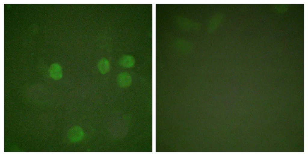
Immunofluorescence analysis of HeLa cells, using Cyclin B1 Antibody. The picture on the right is blocked with the synthesized peptide.
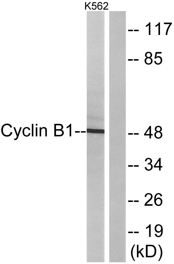
Western blot analysis of lysates from K562 cells, treated with serum 10% 15′, using Cyclin B1 Antibody. The lane on the right is blocked with the synthesized peptide.
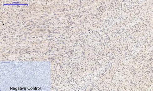
Immunohistochemical analysis of paraffin-embedded Human-uterus tissue. 1,Cyclin B1 Polyclonal Antibody was diluted at 1:200(4°C,overnight). 2, Sodium citrate pH 6.0 was used for antibody retrieval(>98°C,20min). 3,Secondary antibody was diluted at 1:200(room tempeRature, 30min). Negative control was used by secondary antibody only.