
E-cadherin Rabbit Polyclonal Antibody
| Cat Number: | ABN10278 |
|---|---|
| Conjugate: | Unconjugated |
| Size: | 100 ug |
| Concentration: | 1 mg/ml |
| Host: | Rabbit |
| Isotype: | IgG |
| Immunogen: | The antiserum was produced against synthesized peptide derived from human Cadherin. range: 1-50 |
| Reactivity: | Human,Mouse,Rat |
| Applications: | WB 1:500-1:1000; IHC 1:100-1:200; ICC/IF 1:100-1:200; ELISA 1:10000-1:20000 |
| Molecular: | Calculated MW: 97 kDa |
| Purification: | Affinity purification |
| Synonyms: | CDH1; CDHE; UVO; Cadherin-1; CAM120/80; Epithelial cadherin; E-cadherin |
| Background: | Anti-E-cadherin Antibody detects endogenous levels of total E-cadherin protein. |
| Form: | Liquid |
| Buffer: | Liquid in PBS containing 50% glycerol, 0.5% protective protein and 0.02% New type preservative N. |
| Storage: | Aliquot and store at -20°C. Avoid freeze/thaw cycles. |
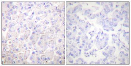
Immunohistochemistry analysis of paraffin-embedded human breast carcinoma tissue, using E-Cadherin Antibody. The picture on the right is Blocked with the synthesized peptide.
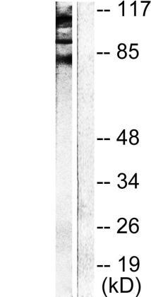
Western blot analysis of lysates from NIH/3T3 cells, using Cadherin Antibody.The lane of the right is blocked with the synthesized peptide.
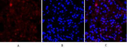
Immunofluorescence analysis of rat-lung tissue. 1,E-cadherin Polyclonal Antibody(red) was diluted at 1:200(4°C,overnight). 2, Cy3 labled Secondary antibody was diluted at 1:300(room temperature, 50min).3, Picture B: DAPI(blue) 10min. Picture A:Target. Picture B: DAPI. Picture C: mergeofA+B