EAAT2 Rabbit Monoclonal Antibody
| Cat Number: | MAB25213 |
|---|---|
| Conjugate: | Unconjugated |
| Size: | 100 ug |
| Clone: | ARC63682 |
| Concentration: | 1mg/ml |
| Host: | Rabbit |
| Isotype: | IgG |
| Immunogen: | Synthetic peptide. This information is considered to be commercially sensitive. |
| Reactivity: | Human,Mouse,Rat |
| Applications: | WB 1:9000 - 1:36000 IHC-P 1:800 - 1:8000 IF/ICC 1:200 - 1:800 ELISA Recommended starting concentration is 1 μg/mL. Please optimize the concentration based on your specific assay requirements. |
| Molecular: | 62kDa/ |
| Purification: | Affinity purification |
| Synonyms: | GLT1; HBGT; DEE41; EAAT2; GLT-1; EIEE41; EAAT2/SLC1A2 |
| Background: | This gene encodes a member of a family of solute transporter proteins. The membrane-bound protein is the principal transporter that clears the excitatory neurotransmitter glutamate from the extracellular space at synapses in the central nervous system. Glutamate clearance is necessary for proper synaptic activation and to prevent neuronal damage from excessive activation of glutamate receptors. Improper regulation of this gene is thought to be associated with several neurological disorders. Alternatively spliced transcript variants of this gene have been identified. |
| Form: | liquid |
| Buffer: | PBS containing 50% glycerol and 0.05% BSA, preserved with proclin300 or sodium azide (as specified on the Certificate of Analysis), pH 7.3. |
| Storage: | Store at -20℃. Avoid freeze / thaw cycles. |

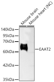
Western blot analysis of various lysates using EAAT2 Rabbit mAbat 1:9000 dilution.
Secondary antibody: HRP-conjugated Goat anti-Rabbit IgG (H+L) at 1:10000 dilution.
Lysates/proteins: 25 μg per lane.
Blocking buffer: 3% nonfat dry milk in TBST.
Detection: ECL Basic Kit.
Negative control (NC): Mouse heart.
Exposure time: 30s.

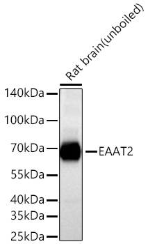
Western blot analysis of lysates from Rat brain using EAAT2 Rabbit mAb at 1:8000 dilution incubated overnight at 4℃.
Secondary antibody: HRP-conjugated Goat anti-Rabbit IgG (H+L) at 1:10000 dilution.
Lysates/proteins: 25 μg per lane.
Blocking buffer: 3% nonfat dry milk in TBST.
Detection: ECL Basic Kit
Exposure time: 10s.

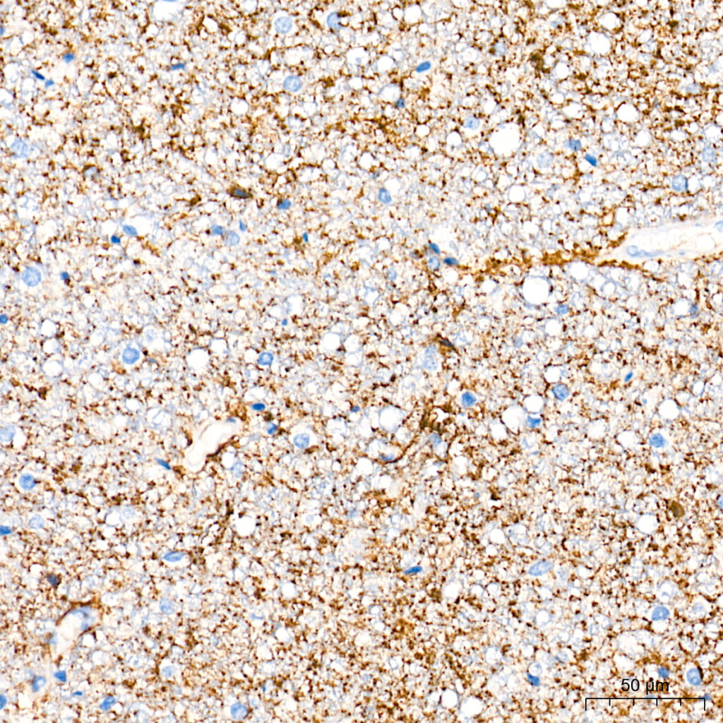
Immunohistochemistry analysis of paraffin-embedded Human brain tissue using EAAT2 Rabbit mAb at a dilution of 1:8000 (40x lens). High pressure antigen retrieval performed with 0.01M Tris-EDTA Buffer(pH 9.0) prior to IHC staining.
