GAP43
| Cat Number: | AB-83908 |
|---|---|
| Conjugate: | Unconjugated |
| Size: | 100 ug |
| Clone: | POLY |
| Concentration: | 1mg/ml |
| Host: | Ch |
| Isotype: | IgY |
| Immunogen: | C-terminal peptide of rodent GAP43, KEDPEADQEHA coupled to KLH |
| Reactivity: | Hu Rt Ms,Cw, Pg, Ho, Ch, |
| Applications: | Western Blot: 1:1,000-1-:5,000 |
| Molecular Weight: | 43kDa by SDS-PAGE |
| Purification: | Purified |
| Background: | GAP43 is an abundant protein which is found heavily concentrated in developing neurons, in particular at the growing tips, the growth cones. One group discovered it since it becomes unregulated during the regeneration of the toad optic nerve, and named it “growth associated protein 43”, the 43 referring to the apparent molecular weight on SDS-PAGE gels (1). GAP43 is very highly charged and does not run on SDS-PAGE in a fashion which accurately reflects its molecular weight, since human GAP43 is 238 amino acids giving a real molecular weight 24.8kDa. The same GAP43 preparation will also give a different SDS-PAGE molecular weight depending on the percentage acrylamide content of the gel, the protein appearing relatively larger on gels with higher acrylamide concentration. GAP43 proteins from different species also may run at different apparent molecular weights on the same gel. Partly due to these unusual features GAP43 was independently discovered by several different groups and therefore has several alternate names, such as protein F1, pp46,neuromodulin, neural phosphoprotein B-50 and calmodulin-binding protein P-57, the numbers 46, 50 and 57 reflecting the apparent SDS-PAGE molecular weight (2). GAP43 is a major protein kinase C substrate and binds calmodulin avidly, this being mediated by an N-terminal IQ calmodulin binding motif (3). GAP43 may be anchored to the plasma membrane by reversible palmitoylation on two Cys residues close to the N-terminus (4). Knock out of the GAP43 gene in mice is lethal early in postnatal life and is associated with defects in axonal pathfinding (5). GAP43 is one of a large family of “intrinsically disordered proteins” which typically have little defined structure unless they are bound to a more structured partner (6). The antibody was made against the full length recombinant human protein and binds to GAP43 in rodents and other mammalian species. It binds strongly to growth cones and axonal processes of neurons in cell culture and to synaptic regions in sectioned material. |
| Form: | Liquid |
| Buffer: | Supplied as an aliquot of IgY preparation plus 5mM NaN3 |
| Storage: | Store at 4°C |

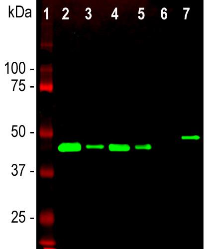
Western blot analysis of different tissue and cell lysates using chicken pAb to GAP43, GAP43, dilution 1:5,000 in green:
[1] protein standard (red),
[2] rat brain,
[3] rat spinal cord,
[4] mouse brain,
[5] mouse spinal cord,
[6] C6 cells,
[7] SH-SY5Y cells.
Single band at the 43kDa mark corresponds to GAP43.The GAP43 protein on ly is detected in the lysates of neuronal origin. C6 cells are a rat glioma cell line and do not express GAP43.

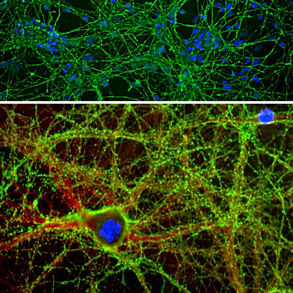
Immunofluorescent analysis of cortical neuron-glial cell culture from E20 rat stained with chicken pAb to GAP43, GAP43, dilution 1:2,000 inb green, and costained with rabbit pAb to α-II spectrin, aII-Spec, dilution 1:1,000 in red. The blue is DAPI staining of nuclear DNA. GAP43 antibody labels protein expressed in the axonal membrane and synapses of neuronal cells, while the spectrin antibody stains the submembraneous
cytoskeleton of axons and dendrites.
