
GluR-1 (phospho Ser863) Rabbit Polyclonal Antibody
| Cat Number: | ABN04735 |
|---|---|
| Conjugate: | Unconjugated |
| Size: | 100ul |
| Clone: | POLY |
| Concentration: | 1mg/ml |
| Host: | Rabbit |
| Isotype: | IgG |
| Immunogen: | Phosphopeptide (Phospho-left) and Non-Phosphopeptide (Phospho-right), using GluR1 (Phospho-Ser863) Antibody Immunohistochemistry analysis of paraffin-embedded human brain, using GluR1 ( Phospho-Ser863) Antibody. The picture on the right is blocked with the phospho peptide. |
| Reactivity: | Human,Mouse,Rat |
| Applications: | WB 1:500-1:2000,IHC 1:100-1:300,ICC/IF 1:50-1:200,ELISA 1:20000-1:40000 |
| Molecular: | 102kDa |
| Purification: | Affinity purification |
| Synonyms: | GRIA1; GLUH1; GLUR1; Glutamate receptor 1; GluR-1; AMPA-selective glutamate receptor 1; GluR-A; GluR-K1; Glutamate receptor ionotropic; AMPA 1; GluA1 |
| Background: | Glutamate receptors are the predominant excitatory neurotransmitter receptors in the mammalian brain and are activated in a variety of normal neurophysiologic processes. These receptors are heteromeric protein complexes with multiple subunits, each possessing transmembrane regions, and all arranged to form a ligand-gated ion channel. The classification of glutamate receptors is based on their activation by different pharmacologic agonists. This gene belongs to a family of alpha-amino-3-hydroxy-5-methyl-4-isoxazole propionate (AMPA) receptors. Alternatively spliced transcript variants encoding different isoforms have been found for this gene. [provided by RefSeq, Jul 2008],function:Ionotropic glutamate receptor. L-glutamate acts as an excitatory neurotransmitter at many synapses in the central nervous system. Binding of the excitatory neurotransmitter L-glutamate induces a conformation change, leading to the opening of the cation channel, and thereby converts the chemical signal to an electrical impulse. The receptor then desensitizes rapidly and enters a transient inactive state, characterized by the presence of bound agonist.,miscellaneous:The postsynaptic actions of Glu are mediated by a variety of receptors that are named according to their selective agonists. This receptor binds AMPA (quisqualate) > glutamate > kainate.,PTM:Palmitoylated. Depalmitoylated upon glutamate stimulation. Cys-603 palmitoylation leads to Golgi retention and decreased cell surface expression. In contrast, Cys-829 palmitoylation does not affect cell surface expression but regulates stimulation-dependent endocytosis.,similarity:Belongs to the glutamate-gated ion channel (TC 1.A.10) family.,subcellular location:Interaction with CACNG2 promotes cell surface expression.,subunit:Homotetramer or heterotetramer of pore-forming glutamate receptor subunits. Tetramers may be formed by the dimerization of dimers. Interacts with DLG1 via its C-terminus. Interacts with CACNG2, HIP1 and RASGRF2.,tissue specificity:Widely expressed in brain., |
| Form: | liquid |
| Buffer: | Liquid in PBS containing 50% glycerol, 0.5% protective protein and 0.02% New type preservative N. |
| Storage: | Store at 4°C short term. Aliquot and store at -20°C long term. Avoid freeze/thaw cycles. |
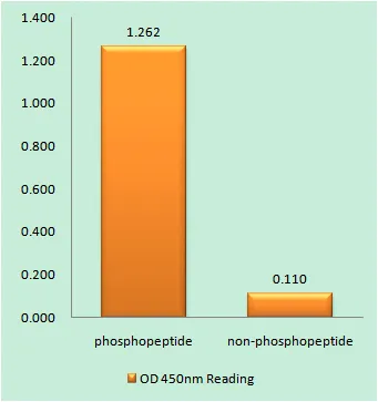
Enzyme-Linked Immunosorbent Assay (Phospho-ELISA) for Immunogen Phosphopeptide (Phospho-left) and Non-Phosphopeptide (Phospho-right), using GluR1 (Phospho-Ser863) Antibody
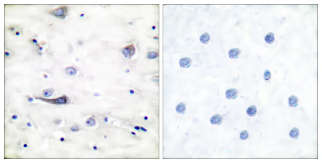
Immunohistochemistry analysis of paraffin-embedded human brain, using GluR1 (Phospho-Ser863) Antibody. The picture on the right is blocked with the phospho peptide.
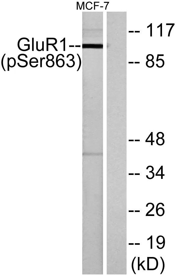
Western blot analysis of lysates from MCF-7 cells, using GluR1 (Phospho-Ser863) Antibody. The lane on the right is blocked with the phospho peptide.