
KD-Validated GPX1 Recombinant Rabbit Monoclonal Antibody
| Cat Number: | MABNK01070 |
|---|---|
| Conjugate: | Unconjugated |
| Size: | 100 ug |
| Host: | Rabbit |
| Isotype: | Rabbit IgG |
| Immunogen: | A synthesized peptide derived from human GPX1. |
| Reactivity: | Human |
| Applications: | WB 1:1,000-1:5,000; FC 1:200-1:2,000; ICC 1:100-1:1,000 |
| Molecular: | Calculated MW: 22.1kDa |
| Purification: | Affinity purification |
| Synonyms: | GPX1; Glutathione Peroxidase 1; Cellular Glutathione Peroxidase; Selenoprotein GPX1; EC 1.11.1.9; GSHPx-1; EC 1.11.1; GSHPX1; GPx-1; GPXD |
| Background: | The protein encoded by this gene belongs to the glutathione peroxidase family, members of which catalyze the reduction of organic hydroperoxides and hydrogen peroxide (H2O2) by glutathione, and thereby protect cells against oxidative damage. Other studies indicate that H2O2 is also essential for growth-factor mediated signal transduction, mitochondrial function, and maintenance of thiol redox-balance; therefore, by limiting H2O2 accumulation, glutathione peroxidases are also involved in modulating these processes. Several isozymes of this gene family exist in vertebrates, which vary in cellular location and substrate specificity. This isozyme is the most abundant, is ubiquitously expressed and localized in the cytoplasm, and whose preferred substrate is hydrogen peroxide. It is also a selenoprotein, containing the rare amino acid selenocysteine (Sec) at its active site. Sec is encoded by the UGA codon, which normally signals translation termination. The 3′ UTRs of selenoprotein mRNAs contain a conserved stem-loop structure, designated the Sec insertion sequence (SECIS) element, that is necessary for the recognition of UGA as a Sec codon, rather than as a stop signal. This gene contains an in-frame GCG trinucleotide repeat in the coding region, and three alleles with 4, 5 or 6 repeats have been found in the human population. The allele with 4 GCG repeats has been significantly associated with breast cancer risk in premenopausal women. Alternatively spliced transcript variants have been found for this gene. Pseudogenes of this locus have been identified on chromosomes X and 21. |
| Form: | Liquid |
| Buffer: | Supplied in PBS (pH 7.4) containing 50% glycerol, and 0.02% sodium azide. |
| Storage: | Aliquot and store at -20°C (valid for 12 months). Avoid freeze/thaw cycles. |
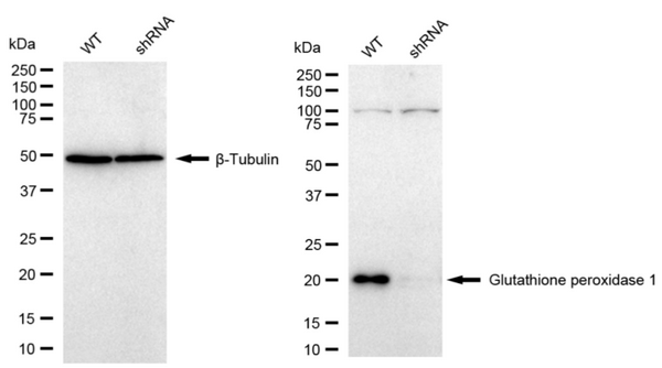
Western blotting analysis using Glutathione peroxidase 1 antibody. Glutathione peroxidase 1 expression in wild type (WT) and Glutathione peroxidase 1 shRNA knockdown (KD) HeLa cells with 30 μg of total cell lysates. β-Tubulin serves as a loading control. The blot was incubated with Glutathione peroxidase 1 antibody ( 1:5,000) and HRP-conjugated goat anti-rabbit secondary antibody (1:10,000) respectively.
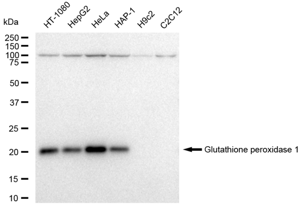
Western blotting analysis using Glutathione peroxidase 1 antibody. Total cell lysates (30 μg) from various cell lines were loaded and separated by SDS-PAGE. The blot was incubated with Glutathione peroxidase 1 antibody (1:5,000) and HRP-conjugated goat anti-rabbit secondary antibody (1:10,000) respectively.
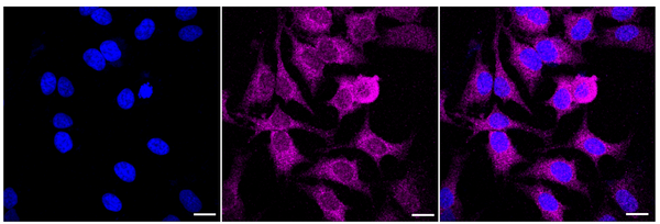
Immunocytochemical staining of Hela cells with Glutathione peroxidase 1 antibody (1:1,000). Nuclei were stained blue with DAPI; Glutathione peroxidase 1 was stained magenta with Alexa Fluor® 647. Images were taken using Leica stellaris 5. Protein abundance based on laser Intensity and smart gain: Low. Scale bar: 20 μm.