
KD-Validated LC3A/LC3B Rabbit Monoclonal Antibody
| Cat Number: | MABNK01267 |
|---|---|
| Conjugate: | Unconjugated |
| Size: | 100 ug |
| Clone: | D3U4C |
| Host: | Rabbit |
| Isotype: | Rabbit IgG |
| Immunogen: | A synthesized peptide derived from human LC3B |
| Reactivity: | Human,Mouse,Rat |
| Applications: | WB 1:1,000-1:5,000; FC 1:200-1:2,000; ICC 1:100-1:1,000; IHC-P 1:100-1:200 |
| Molecular: | Calculated MW: 14.7kDa |
| Purification: | Affinity purification |
| Synonyms: | MAP1LC3BMicrotubule-Associated Proteins 1A/1B Light Chain 3B; Autophagy-Related Ubiquitin-Like Modifier LC3 B; MAP1 Light Chain 3-Like Protein 2; MAP1A/MAP1B Light Chain 3 B; MAP1A/MAP1B LC3 B; MAP1A/1BLC3; MAP1LC3B-A; MAP1ALC3; LC3B |
| Background: | The product of this gene is a subunit of neuronal microtubule-associated MAP1A and MAP1B proteins, which are involved in microtubule assembly and important for neurogenesis. Studies on the rat homolog implicate a role for this gene in autophagy, a process that involves the bulk degradation of cytoplasmic component. [provided by RefSeq, Jul 2008] |
| Form: | Liquid |
| Buffer: | Supplied in PBS (pH 7.4) containing 50% glycerol, and 0.02% sodium azide. |
| Storage: | Aliquot and store at -20°C (valid for 12 months). Avoid freeze/thaw cycles. |
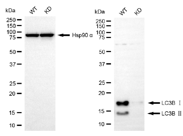
Western blotting analysis usingLC3A/LC3B antibody. LC3B expression in wild-type (WT) and LC3A/LC3B knockdown (KD) HeLa cells with 20 μg of total cell lysates. Hsp90 α serves as a loading control. The blot was incubated with LC3B antibody (1:5,000) and HRP-conjugated goat anti-rabbit secondary antibody (1:10,000) respectively.
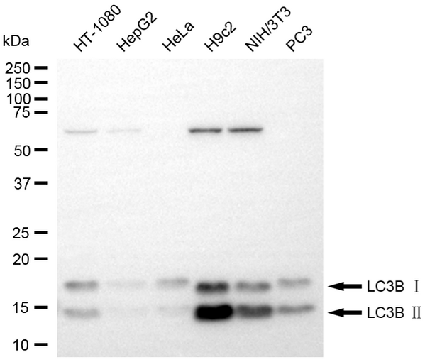
Western blotting analysis using LC3A/LC3B antibody . Total cell lysates (30 μg) from various cell lines were loaded and separated by SDS-PAGE. The blot was incubated with LC3B antibody 1:5,000) and HRP-conjugated goat anti-rabbit secondary antibody (1:10,000) respectively.
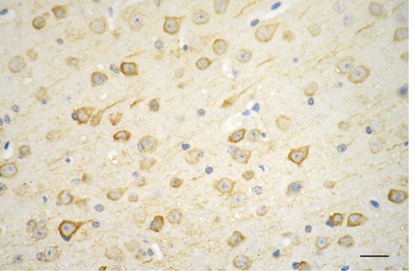
Immunohistochemistry was performed on paraffin-embedded mouse brain using LC3A/LC3B antibody (1:200). Antigen retrieval was done in sodium citrate buffer (pH 6.0). DAB was used for detection, with hematoxylin counterstaining. Images were acquired using a Nikon Ci-L Plus microscope (40× objective). Scale bar: 25 μm.