LAMP2 / CD107b
| Cat Number: | MAB-94429 |
|---|---|
| Conjugate: | Unconjugated |
| Size: | 100 μg |
| Clone: | H4B4 |
| Concentration: | 1mg/ml |
| Host: | Mouse |
| Isotype: | IgG1 kappa |
| Immunogen: | Human PBMC |
| Reactivity: | Human |
| Applications: | Flow cytometry: Intracellular and extracellular staining; recommended dilution: 1-2 μg/ml. Immunohistochemistry: Recommended dilution: 2-8 μg/ml. Western blotting: Recommended dilution: 1-2 μg/ml. |
| Molecular: | 100-120kDa |
| Purification: | Purified by protein-A affinity chromatography |
| Synonyms: | LAMP-2, LAMPB |
| Background: | CD107b (lysosome-associated membrane protein-2, LAMP-2), together with CD107a / LAMP-1, is a major constituent of lysosomal membrane. The LAMP proteins are involved in lysosome biogenesis and are required for fusion of lysosomes with phagosomes, especially CD107b is important regulator in successful phagosomal maturation. CD107b deficiency causes an accumulation of autophagosomes in many tissues leading to cardiomyopathy and myopathy (Danons disease). Immature CD107b is an approximately 45 kDa protein, but after extensive glycosylation the mature glycoprotein has about 100-120 kDa |
| Form: | Liquid |
| Buffer: | Phosphate buffered saline (PBS), pH 7.4 |
| Storage: | Store at 2-8°C. Do not freeze. |
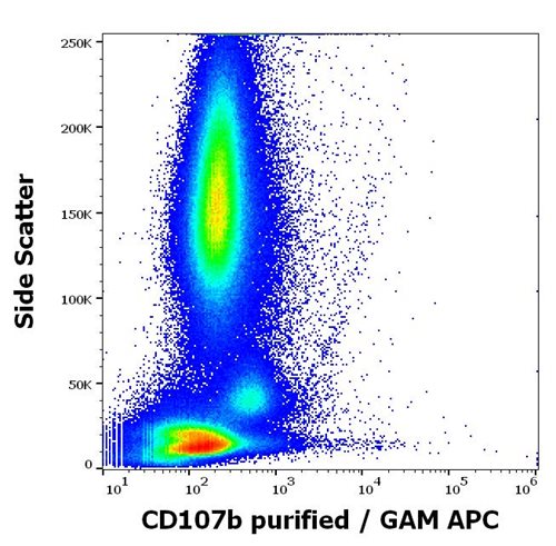
Flow cytometry surface staining pattern of human anti-IgE antibody stimulated peripheral whole blood stained using anti-human CD107b (H4B4) purified antibody (concentration in sample 1,67 μg/ml, GAM APC)
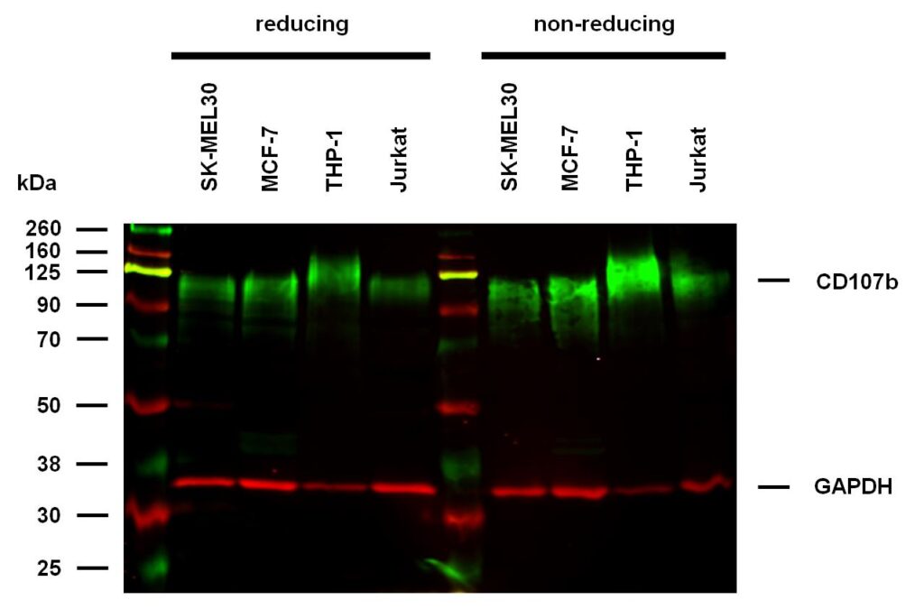
Anti-Hu CD107b Purified (clone H4B4) works in WB application under reducing and non-reducing conditions.
Western blotting analysis was performed on whole cell extracts (RIPA lysis buffer) of SK-MEL30, MCF-7, THP-1, and Jurkat cell lines, mixed and heated (100°C, 5 min) with reducing and non-reducing SDS-loading buffer. Samples were resolved using 10% SDS-PAGE gel.
Nitrocellulose membrane blot was probed with mouse IgG1 monoclonal antibody H4B4 (1 µg/ml), followed by IRDye 800CW Goat-anti-Mouse IgG (green). Mouse anti-GAPDH monoclonal antibody FF26A conjugated with DyLight 680 (0.1 µg/ml) was used as the loading control (red). Multiplex fluorescent Western blot detection was performed.
CD107b molecules were detected at ~95-125 kDa in all tested cell lines under both reducing and non-reducing conditions.