LAMP3 /CD63 Mouse Monoclonal Antibody
| Cat Number: | MAB-10067 |
|---|---|
| Conjugate: | Unconjugated |
| Size: | 100 ug |
| Clone: | MEM-259 |
| Concentration: | 1mg/ml |
| Host: | Mouse |
| Isotype: | IgG1 |
| Immunogen: | HPB-ALL T cell line |
| Reactivity: | Human |
| Applications: | Flow cytometry: Recommended dilution: 2 μg/ml; positive material: activated platelets, neutrophils and basophils. Immunohistochemistry (paraffin sections): Recommended dilution: 10 μg/ml; positive tissue: spleen. |
| Molecular: | 40-60 kDa |
| Purification: | Purified by protein-A affinity chromatography. |
| Synonyms: | OMA81H, Granulophysin, Tetraspanin-30, Tspan-30, MLA1, ME491, LAMP-3, OMA81H, TSPAN30 |
| Background: | CD63 (LAMP-3, lysosome-associated membrane protein-3), a glycoprotein of tetraspanin family, is present in late endosomes, lysosomes and secretory vesicles of various cell types. It is also present in the plasma membrane, usually following cell activation. Hence, it has become an widely used basophil activation marker. In mast cells, however, CD63 exposition does not need their activation. CD63 interacts with integrins and affects phagocytosis and cell migration, it is also involved in H/K-ATPase trafficking regulation of ROMK1 channels. CD63 also serves as a T-cell costimulation molecule. Expression of CD63 can be used for predicting the prognosis in earlier stages of carcinomas |
| Form: | Liquid |
| Buffer: | Phosphate buffered saline (PBS), pH 7.4, 15 mM sodium azide |
| Storage: | Store at 2-8°C. Do not freeze. |
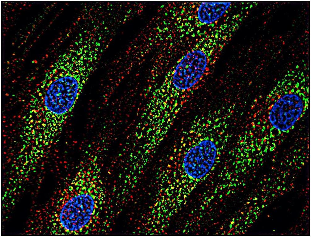
Immunocytochemistry staining of human skin fibroblasts with anti-CD63 (MEM-259; green) after co-incubation of living cells with human Transferrin – 547 cell nuclei stained with DAPI (blue)

Immunohistochemistry staining of human spleen (paraffin sections) using anti-CD63 .
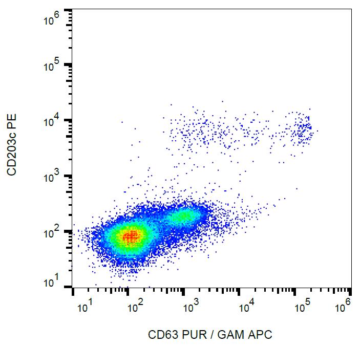
Flow cytometry analysis of IgE-activated peripheral blood stained with anti-human CD63 (MEM-259) purified, GAM-APC.