NFKB1 Rabbit Monoclonal antibody
| Cat Number: | AB27463 |
|---|---|
| Conjugate: | Unconjugated |
| Size: | 100 ug |
| Concentration: | 1mg/ml |
| Host: | Rabbit |
| Isotype: | IgG |
| Immunogen: | Synthetic peptide. This information is considered to be commercially sensitive. |
| Reactivity: | Human,Mouse |
| Applications: | WB 1:1000 - 1:2000 IHC-P 1:200 - 1:400 IP 0.5μg-4μg antibody for 200μg-400μg extracts of whole cells ELISA Recommended starting concentration is 1 μg/mL ChIP 5μg antibody for 5μg-20μg of Chromatin. Please optimize the concentration based on your specific assay requirements. |
| Molecular: | 50 kDa(Active form)/120 kDa(Precursor) |
| Purification: | Affinity purification |
| Synonyms: | KBF1; EBP-1; NF-kB; CVID12; NF-kB1; NFKB-p50; NFkappaB; NF-kappaB; NFKB-p105; NF-kappa-B1; NF-kappabeta |
| Background: | This gene encodes a 105 kD protein which can undergo cotranslational processing by the 26S proteasome to produce a 50 kD protein. The 105 kD protein is a Rel protein-specific transcription inhibitor and the 50 kD protein is a DNA binding subunit of the NF-kappa-B (NFKB) protein complex. NFKB is a transcription regulator that is activated by various intra- and extra-cellular stimuli such as cytokines, oxidant-free radicals, ultraviolet irradiation, and bacterial or viral products. Activated NFKB translocates into the nucleus and stimulates the expression of genes involved in a wide variety of biological functions. Inappropriate activation of NFKB has been associated with a number of inflammatory diseases while persistent inhibition of NFKB leads to inappropriate immune cell development or delayed cell growth. NFKB is a critical regulator of the immediate-early response to viral infection. Alternative splicing results in multiple transcript variants encoding different isoforms, at least one of which is proteolytically processed. |
| Form: | liquid |
| Buffer: | PBS containing 50% glycerol and 0.05% BSA, preserved with proclin300 or sodium azide (as specified on the Certificate of Analysis), pH 7.3. |
| Storage: | Store at -20℃. Avoid freeze / thaw cycles. |
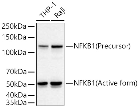
Western blot analysis of various lysates using NFKB1 Rabbit mAb at 1:1000 dilution incubated overnight at 4℃.
Secondary antibody: HRP-conjugated Goat anti-Rabbit IgG (H+L) at 1:10000 dilution.
Lysates/proteins: 25 μg per lane.
Blocking buffer: 3% nonfat dry milk in TBST.
Detection: ECLWest Pico Plus.
Exposure time: 10 s.
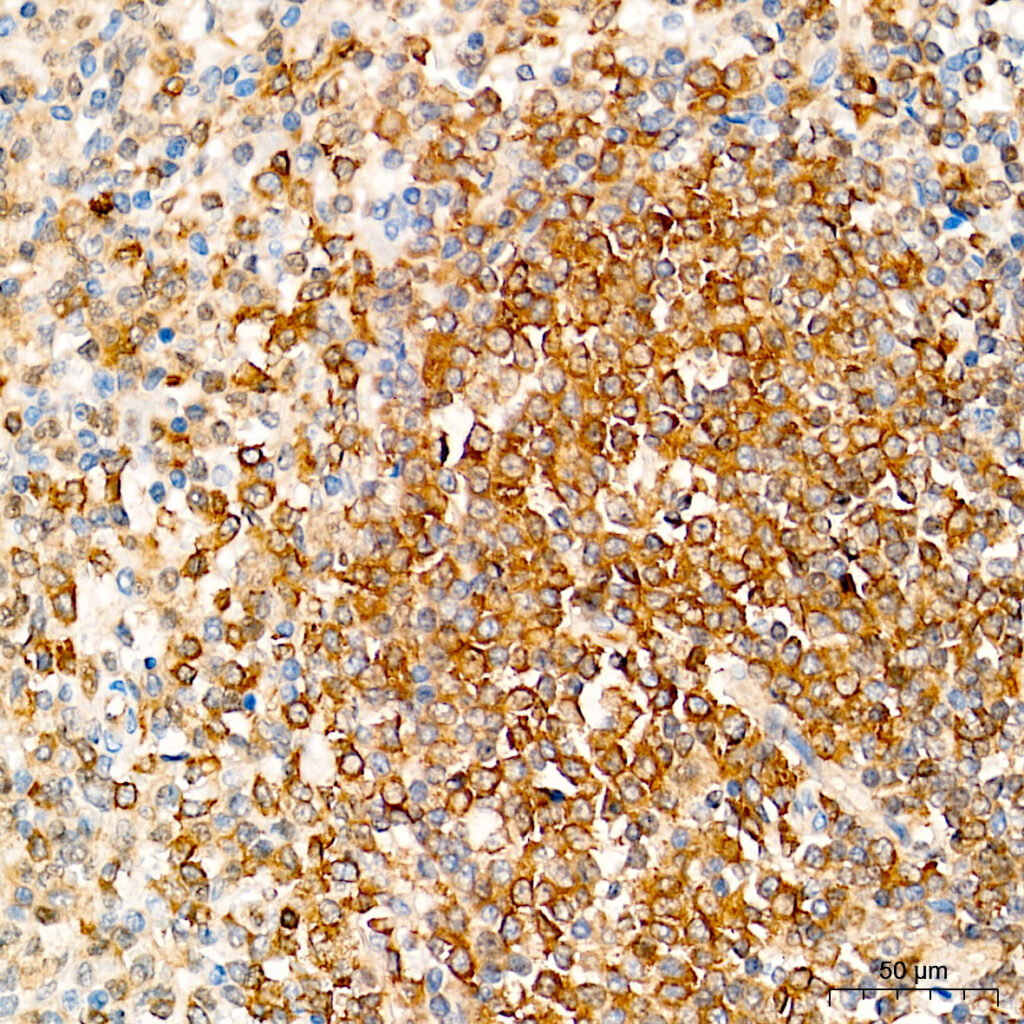
Immunohistochemistry analysis of paraffin-embedded Human spleen tissue using NFKB1 Rabbit mAb at a dilution of 1:200 (40x lens). High pressure antigen retrieval performed with 0.01M Tris-EDTA Buffer (pH 9.0) prior to IHC staining.
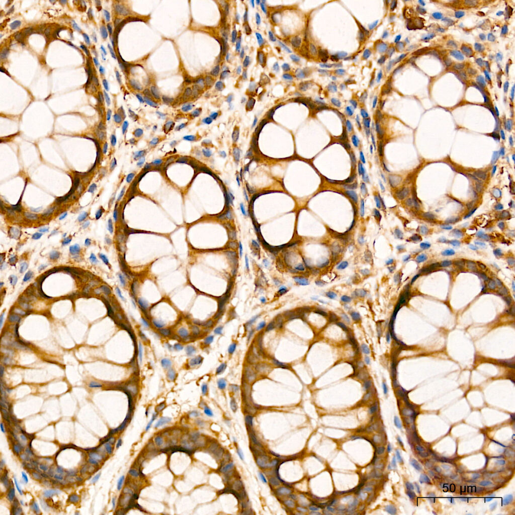
Immunohistochemistry analysis of paraffin-embedded Human colon tissue using NFKB1 Rabbit mAb at a dilution of 1:200 (40x lens). High pressure antigen retrieval performed with 0.01M Tris-EDTA Buffer (pH 9.0) prior to IHC staining.