NG2/CSPG4 Rabbit Monoclonal Antibody
| Cat Number: | MAB-94805 |
|---|---|
| Conjugate: | Unconjugated |
| Size: | 100 ug |
| Clone: | ARC64580 |
| Concentration: | 1mg/ml |
| Host: | Rabbit |
| Isotype: | IgG |
| Immunogen: | Recombinant protein.This information is considered to be commercially sensitive. |
| Reactivity: | Human,Mouse |
| Applications: | WB 1:1000 - 1:2000 IF-P 1:200 - 1:1000 IHC-P 1:500 - 1:2000 FC 1:500 - 1:1000 ELISA Recommended starting concentration is 1 μg/mL. Please optimize the concentration based on your specific assay requirements. |
| Molecular: | 450kDa |
| Purification: | Affinity purification |
| Synonyms: | NG2; MCSP; MCSPG; MSK16; CSPG4A; HMW-MAA; MEL-CSPG; NG2/CSPG4 |
| Background: | A human melanoma-associated chondroitin sulfate proteoglycan plays a role in stabilizing cell-substratum interactions during early events of melanoma cell spreading on endothelial basement membranes. CSPG4 represents an integral membrane chondroitin sulfate proteoglycan expressed by human malignant melanoma cells. |
| Form: | liquid |
| Buffer: | PBS containing 50% glycerol and 0.05% BSA, preserved with proclin300 or sodium azide (as specified on the Certificate of Analysis), pH 7.3. |
| Storage: | Store at -20℃. Avoid freeze / thaw cycles. |
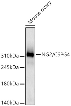
Western blot analysis of lysates from Mouse ovary using NG2/CSPG4 Rabbit mAb at 1:1000 dilution incubated overnight at 4℃.
Secondary antibody: HRP-conjugated Goat anti-Rabbit IgG (H+L) at 1:10000 dilution.
Lysates/proteins: 25 μg per lane.
Blocking buffer: 3% nonfat dry milk in TBST.
Detection: ECL West Pico Plus.
Exposure time: 60s.
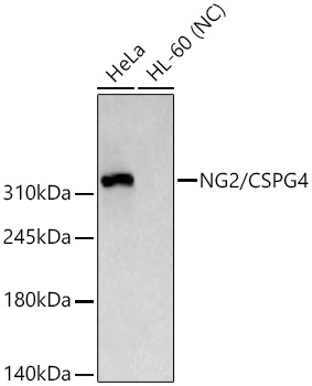
Western blot analysis of various lysates using NG2/CSPG4 Rabbit mAb at 1:1000 dilution incubated overnight at 4℃.
Secondary antibody: HRP-conjugated Goat anti-Rabbit IgG (H+L) at 1:10000 dilution.
Lysates/proteins: 25 μg per lane.
Blocking buffer: 3% nonfat dry milk in TBST.
Detection: ECL West Pico Plus.
Negative control (NC): HL-60
Exposure time: 10s.
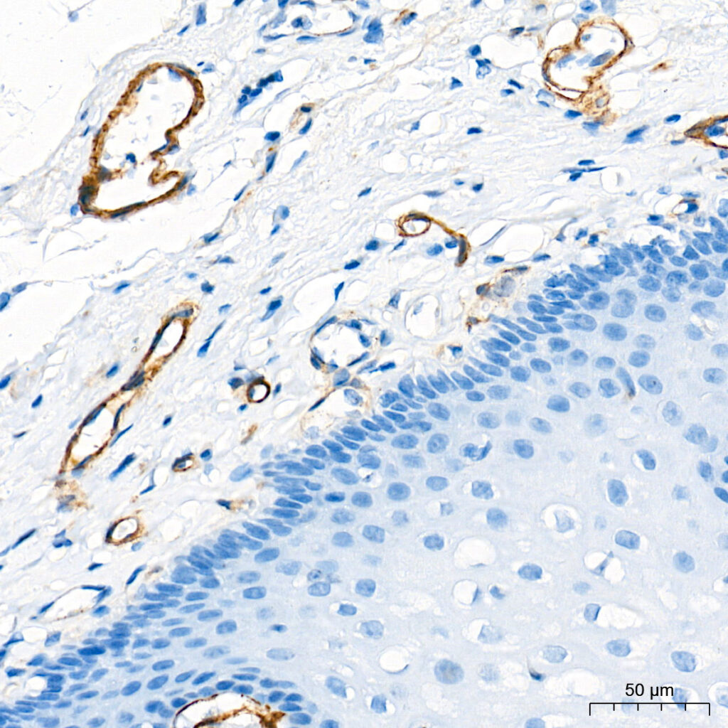
Immunohistochemistry analysis of paraffin-embedded Human esophagus tissue using NG2/CSPG4 Rabbit mAb at a dilution of 1:1000 (40x lens). High pressure antigen retrieval performed with 0.01M Tris-EDTA Buffer (pH 9.0) prior to IHC staining.