NLRP3 Rabbit Monoclonal Antibody
| Cat Number: | MAB24294 |
|---|---|
| Conjugate: | Unconjugated |
| Size: | 100 ug |
| Clone: | ARC62768 |
| Concentration: | 1mg/ml |
| Host: | Rabbit |
| Isotype: | IgG |
| Immunogen: | Recombinant protein.This information is considered to be commercially sensitive. |
| Reactivity: | Mouse |
| Applications: | WB 1:1000 - 1:6000 IF/ICC 1:100 - 1:500 IP 0.5μg-4μg antibody for 200μg-400μg extracts of whole cells ELISA Recommended starting concentration is 1 μg/mL. Please optimize the concentration based on your specific assay requirements. |
| Molecular: | 118kDa |
| Purification: | Affinity purification |
| Synonyms: | FCU; MWS; FCAS; Cias1; Mmig1; NALP3; Pypaf1; AII/AVP; AGTAVPRL; NLRP3 |
| Background: | Enables DNA-binding transcription factor binding activity and sequence-specific DNA binding activity. Involved in several processes, including positive regulation of T-helper cell differentiation; positive regulation of cytokine production; and response to bacterium. Acts upstream of or within several processes, including NLRP3 inflammasome complex assembly; activation of cysteine-type endopeptidase activity involved in apoptotic process; and defense response to virus. Located in cytoplasm and nucleus. Part of NLRP3 inflammasome complex. Is expressed in central nervous system and retina. Used to study CINCA Syndrome; familial cold autoinflammatory syndrome 1; and non-alcoholic fatty liver disease. Human ortholog(s) of this gene implicated in CINCA Syndrome; Muckle-Wells syndrome; autosomal dominant nonsyndromic deafness 34; familial cold autoinflammatory syndrome 1; and urticaria. Orthologous to human NLRP3 (NLR family pyrin domain containing 3). |
| Form: | liquid |
| Buffer: | PBS with 0.09% Sodium azide,0.05% BSA,50% glycerol,pH7.3. |
| Storage: | Store at -20℃. Avoid freeze / thaw cycles. |
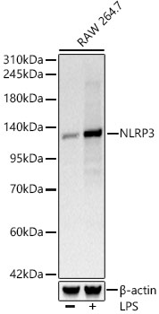
Western blot analysis of lysates from RAW 264.7 cells, using NLRP3 Rabbit mAb at 1:1000 dilution. Raw264. 7 cells were treated with LPS (1 μg/ml) at 37℃ for 8 hours.
Secondary antibody: HRP-conjugated Goat anti-Rabbit IgG (H+L) ) at 1:10000 dilution.
Lysates/proteins: 25μg per lane.
Blocking buffer: 3% nonfat dry milk in TBST.
Detection: ECL West Pico Plus.
Exposure time: 10s.
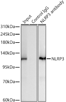
Immunoprecipitation of NLRP3 from 300 µg extracts of RAW 264.7 cells treated with LPS (1 μg/ml, 8h) was performed using 3 µg of NLRP3 Rabbit mAb ). Rabbit IgG isotype control was used to precipitate the Control IgG sample. IP samples were eluted with 1X Laemmli Buffer. The Input lane represents 10% of the total input. Western blot analysis of immunoprecipitates was conducted using NLRP3 Rabbit mAb at a dilution of 1:2000.