PD-L1 (CD274) Rabbit Polyclonal Antibody
| Cat Number: | AB-84131 |
|---|---|
| Conjugate: | Unconjugated |
| Size: | 100ug |
| Clone: | POLY |
| Concentration: | 1mg/ml |
| Host: | Rabbit |
| Isotype: | IgG |
| Immunogen: | Recombinant fusion protein containing a sequence corresponding to amino acids 131-239 of human PD-L1/CD274 |
| Reactivity: | Human, Mouse |
| Applications: | Western Blot: 1:500 - 1:1000Immunohistochemistry: 1:50 - 1:200 |
| Molecular: | 40KDa |
| Purification: | Aff. Pur. |
| Synonyms: | CD274; B7-H; B7H1; PD-L1; PDCD1L1; PDCD1LG1; PDL1; CD274 molecule |
| Background: | This gene encodes an immune inhibitory receptor ligand that is expressed by hematopoietic and non-hematopoietic cells, such as T cells and B cells and various types of tumor cells. The encoded protein is a type I transmembrane protein that has immunoglobulin V-like and C-like domains. Interaction of this ligand with its receptor inhibits T-cell activation and cytokine production. During infection or inflammation of normal tissue, this interaction is important for preventing autoimmunity by maintaining homeostasis of the immune response. In tumor microenvironments, this interaction provides an immune escape for tumor cells through cytotoxic T-cell inactivation. |
| Form: | Liquid |
| Buffer: | PBS with 0.01% thiomersal,50% glycerol,pH7.3. |
| Storage: | Store at -20℃. Avoid freeze / thaw cycles. |
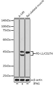
Western blot analysis of various lysates using PD-L1/CD274 Rabbit pAb (A11273) at
1:500 dilution. A-549 cells were treated by IFNG (100 ng/mL) at 37℃ for 48 hours.
Secondary antibody: HRP-conjugated Goat anti-Rabbit IgG (H+L) (AS014) at 1:10000
dilution.
Lysates/proteins: 25μg per lane.
Blocking buffer: 3% nonfat dry milk in TBST.
Detection: ECL Basic Kit (RM00020).
Exposure time: 180s.