
PDX1 (9B19) Rabbit Monoclonal Antibody
| Cat Number: | MABN15935 |
|---|---|
| Conjugate: | Unconjugated |
| Size: | 100 ug |
| Clone: | 9B19 |
| Concentration: | 1mg/ml. The concentration of this product may be batch-dependent. |
| Host: | Rabbit |
| Isotype: | IgG |
| Immunogen: | Recombinant protein of human PDX1 |
| Reactivity: | Human,Mouse,Rat |
| Applications: | WB 1:500-1:2000, IHC 1:200-1:500, ICC/IF 1:500-1:1000, IP 1:20-1:50 |
| Molecular: | 31 kDa |
| Purification: | Affinity purification |
| Synonyms: | GSF;IPF1;IUF1;IDX-1;MODY4;PDX-1;STF-1 |
| Background: | The protein encoded by this gene is a transcriptional activator of several genes, including insulin, somatostatin, glucokinase, islet amyloid polypeptide, and glucose transporter type 2. The encoded nuclear protein is involved in the early development of the pancreas and plays a major role in glucose-dependent regulation of insulin gene expression. Activates insulin, somatostatin, glucokinase, islet amyloid polypeptide and glucose transporter type 2 gene transcription. Particularly involved in glucose-dependent regulation of insulin gene transcription. As part of a PDX1:PBX1b:MEIS2b complex in pancreatic acinar cells is involved in the transcriptional activation of the ELA1 enhancer; the complex binds to the enhancer B element and cooperates with the transcription factor 1 complex (PTF1) bound to the enhancer A element. Binds preferentially the DNA motif 5′-[CT]TAAT[TG]-3′. During development, specifies the early pancreatic epithelium, permitting its proliferation, branching and subsequent differentiation. At adult stage, required for maintaining the hormone-producing phenotype of the beta-cell. |
| Form: | Liquid |
| Buffer: | Rabbit IgG in phosphate buffered saline, pH 7.4, 150mM NaCl, 0.02% New type preservative N and 50% glycerol. Store at +4°C short term. Store at -20°C long term. Avoid freeze/thaw cycle. |
| Storage: | Aliquot and store at -20°C (valid for 12 months). Avoid freeze/thaw cycles. |
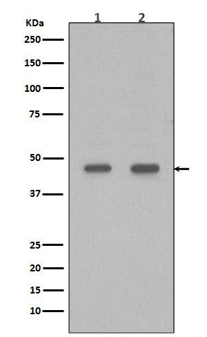
Western blot analysis of PDX1 in (1) Caco-2 cell Lysate; (2) BxPC-3 cell Lysate.
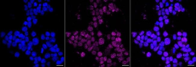
Immunocytochemistry analysis of PDX1 in HeLa cells. Sample was incubated with PDX1 monoclonal antibody at a dilution of 1:1,000, followed by DAPI and Alexa Fluor 647. Protein abundance based on laser intensity and smart gain: Medium. Scale bar, 20μm.
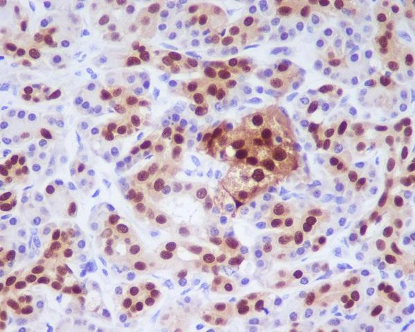
Immunohistochemical (Paraffin) analysis of PDX1 in Human pancreas using PDX1 Recombinant Monoclonal Antibody. High-pressure and temperature Sodium Citrate pH 6.0 was used for antigen retrieval.