PGK1 Rabbit pAb
| Cat Number: | AB14039 |
|---|---|
| Conjugate: | Unconjugated |
| Size: | 100 ug |
| Concentration: | 1mg/ml |
| Host: | Rabbit |
| Isotype: | IgG |
| Immunogen: | Recombinant protein.This information is considered to be commercially sensitive. |
| Reactivity: | Human,Mouse,Rat |
| Applications: | WB 1:100 - 1:500 IHC-P 1:50 - 1:200 ELISA Recommended starting concentration is 1 μg/mL. Please optimize the concentration based on your specific assay requirements. |
| Molecular: | 45kDa |
| Purification: | Affinity purification |
| Synonyms: | PGKA; MIG10; HEL-S-68p; PGK1 |
| Background: | The protein encoded by this gene is a glycolytic enzyme that catalyzes the conversion of 1,3-diphosphoglycerate to 3-phosphoglycerate. The encoded protein may also act as a cofactor for polymerase alpha. Additionally, this protein is secreted by tumor cells where it participates in angiogenesis by functioning to reduce disulfide bonds in the serine protease, plasmin, which consequently leads to the release of the tumor blood vessel inhibitor angiostatin. The encoded protein has been identified as a moonlighting protein based on its ability to perform mechanistically distinct functions. Deficiency of the enzyme is associated with a wide range of clinical phenotypes hemolytic anemia and neurological impairment. Pseudogenes of this gene have been defined on chromosomes 19, 21 and the X chromosome. |
| Form: | liquid |
| Buffer: | PBS with 0.09% Sodium azide,50% glycerol,pH7.3. |
| Storage: | Store at -20℃. Avoid freeze / thaw cycles. |
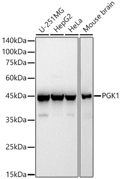
Western blot analysis of various lysates, using PGK1 Rabbit pAb at 1:400 dilution.
Secondary antibody: HRP-conjugated Goat anti-Rabbit IgG (H+L) (AS014) at 1:10000 dilution.
Lysates/proteins: 25μg per lane.
Blocking buffer: 3% nonfat dry milk in TBST.
Detection: ECL Basic Kit (RM00020).
Exposure time: 60s.
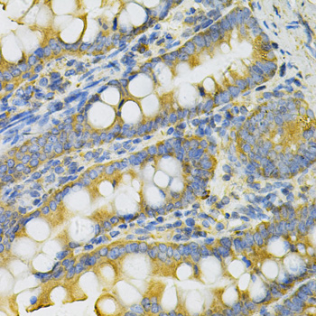
Immunohistochemistry analysis of paraffin-embedded Rat intestine using PGK1 Rabbit pAb at dilution of 1:100 (40x lens). Microwave antigen retrieval performed with 0.01M PBS Buffer (pH 7.2) prior to IHC staining.
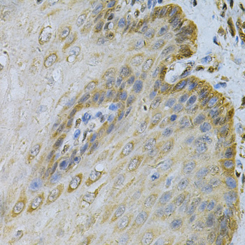
Immunohistochemistry analysis of paraffin-embedded Human esophagus using PGK1 Rabbit pAb at dilution of 1:100 (40x lens). Microwave antigen retrieval performed with 0.01M PBS Buffer (pH 7.2) prior to IHC staining.