Phospho-Akt-S473 Rabbit mAb
| Cat Number: | MAB1453 |
|---|---|
| Conjugate: | Unconjugated |
| Size: | 100 ug |
| Clone: | ARC5023-02 |
| Concentration: | 1mg/ml |
| Host: | Rabbit |
| Isotype: | IgG |
| Immunogen: | A synthetic phosphorylated peptide around S473 of human AKT1 (NP_005154.2). |
| Reactivity: | Human,Mouse,Rat |
| Applications: | WB,1:500 - 1:1000 IHC-P,1:50 - 1:200 ELISA,Recommended starting concentration is 1 μg/mL. Please optimize the concentration based on your specific assay requirements. |
| Molecular: | 60kDa/ |
| Purification: | Affinity purification |
| Background: | The serine-threonine protein kinase encoded by the AKT1 gene is catalytically inactive in serum-starved primary and immortalized fibroblasts. AKT1 and the related AKT2 are activated by platelet-derived growth factor. The activation is rapid and specific, and it is abrogated by mutations in the pleckstrin homology domain of AKT1. It was shown that the activation occurs through phosphatidylinositol 3-kinase. In the developing nervous system AKT is a critical mediator of growth factor-induced neuronal survival. Survival factors can suppress apoptosis in a transcription-independent manner by activating the serine/threonine kinase AKT1, which then phosphorylates and inactivates components of the apoptotic machinery. Mutations in this gene have been associated with the Proteus syndrome. Multiple alternatively spliced transcript variants have been found for this gene. [provided by RefSeq, Jul 2011] |
| Form: | liquid |
| Buffer: | PBS with 0.09% sodium azide,0.05% BSA,50% glycerol,pH7.3. |
| Storage: | Store at -20℃. Avoid freeze / thaw cycles. |
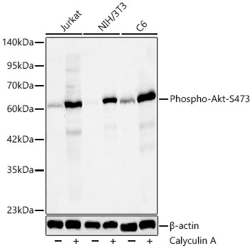
Western blot analysis of lysates from Jurkat,NIH/3T3,C6 cells using Phospho-Akt-S473 Rabbit mAb at 1:1000 dilution. Jurkat,NIH/3T3 and C6 cells were treated by Calyculin A at 37℃ for 30 minutes.
Secondary antibody: HRP-conjugated Goat anti-Rabbit IgG at 1:10000 dilution.
Lysates/proteins: 25 μg per lane.
Blocking buffer: 3% nonfat dry milk in TBST.
Detection: ECL Basic Kit .
Exposure time: 20s.
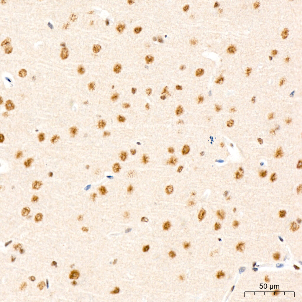
Immunohistochemistry analysis of paraffin-embedded Mouse brain tissue using Phospho-Akt-S473 Rabbit mAb at a dilution of 1:200 . High pressure antigen retrieval performed with 0.01M Citrate Bufferr prior to IHC staining.
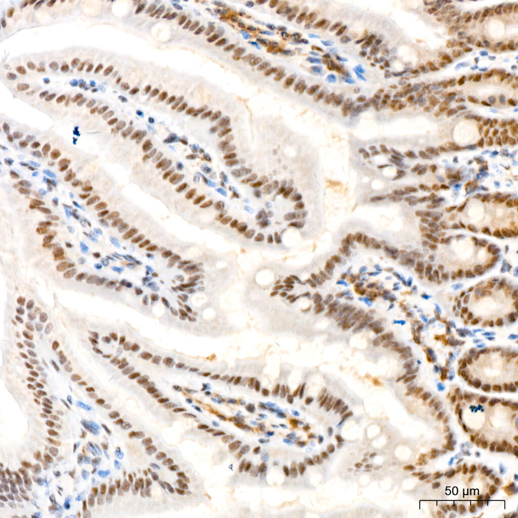
Immunohistochemistry analysis of paraffin-embedded Mouse colon tissue using Phospho-Akt-S473 Rabbit mAb at a dilution of 1:200 . High pressure antigen retrieval performed with 0.01M Citrate Bufferr prior to IHC staining.