S100B Rabbit Polyclonal Antibody
| Cat Number: | AB0676 |
|---|---|
| Conjugate: | Unconjugated |
| Size: | 100 ug |
| Concentration: | 1mg/ml |
| Host: | Rabbit |
| Isotype: | IgG |
| Immunogen: | Recombinant protein.This information is considered to be commercially sensitive. |
| Reactivity: | Human,Mouse,Rat |
| Applications: | WB 1:500 - 1:1000 IP 0.5μg-4μg antibody for 400μg-600μg extracts of whole cells IF-P 1:50 - 1:200 IHC-P 1:50 - 1:200 ELISA Recommended starting concentration is 1 μg/mL. Please optimize the concentration based on your specific assay requirements. |
| Molecular: | 11kDa |
| Purification: | Affinity purification |
| Synonyms: | NEF; S100; S100-B; S100beta; S100B |
| Background: | The protein encoded by this gene is a member of the S100 family of proteins containing 2 EF-hand calcium-binding motifs. S100 proteins are localized in the cytoplasm and/or nucleus of a wide range of cells, and involved in the regulation of a number of cellular processes such as cell cycle progression and differentiation. S100 genes include at least 13 members which are located as a cluster on chromosome 1q21; however, this gene is located at 21q22.3. This protein may function in Neurite extension, proliferation of melanoma cells, stimulation of Ca2+ fluxes, inhibition of PKC-mediated phosphorylation, astrocytosis and axonal proliferation, and inhibition of microtubule assembly. Chromosomal rearrangements and altered expression of this gene have been implicated in several neurological, neoplastic, and other types of diseases, including Alzheimer’s disease, Down’s syndrome, epilepsy, amyotrophic lateral sclerosis, melanoma, and type I diabetes. |
| Form: | liquid |
| Buffer: | PBS with 0.09% Sodium azide,50% glycerol,pH7.3. |
| Storage: | Store at -20℃. Avoid freeze / thaw cycles. |
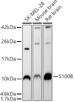
Western blot analysis of various lysates using S100B Rabbit pAb at 1:500 dilution.
Secondary antibody: HRP-conjugated Goat anti-Rabbit IgG (H+L)at 1:10000 dilution.
Lysates / proteins: 25 μg per lane.
Blocking buffer: 3 % nonfat dry milk in TBST.
Detection: ECL West Pico Plus.
Exposure time: 90s.
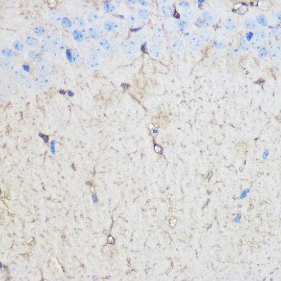
Immunohistochemistry analysis of paraffin-embedded Mouse brain using S100B Rabbit pAb at dilution of 1:100 (40x lens). Microwave antigen retrieval performed with 0.01M PBS Buffer (pH 7.2) prior to IHC staining.
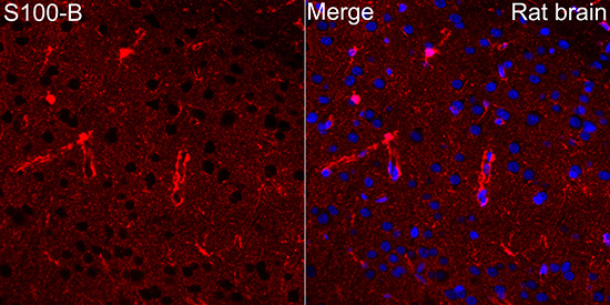
Immunofluorescence analysis of paraffin-embedded rat brain using S100B Rabbit pAb at dilution of 1:50 (40x lens). Secondary antibody: Cy3-conjugated Goat anti-Rabbit IgG (H+L) at 1:500 dilution. Blue: DAPI for nuclear staining. Perform microwave antigen retrieval with 0.01M citrate buffer (pH 6.0) prior to IF staining.