
SQSTM1/p62 Rabbit Monoclonal Antibody
| Cat Number: | MAB19700 |
|---|---|
| Conjugate: | Unconjugated |
| Size: | 100 ug |
| Clone: | ARC0180 |
| Host: | Rabbit |
| Isotype: | IgG |
| Immunogen: | Synthetic peptide. This information is considered to be commercially sensitive. |
| Reactivity: | Human,Mouse,Rat |
| Applications: | WB 1:20000 - 1:80000; IHC-P 1:500 - 1:5000; IF/ICC 1:50 - 1:200; IP 0.5μg-4μg antibody for 200μg-400μg extracts of whole cells; ELISA Recommended starting concentration is 1 μg/mL. Please optimize the concentration based on your specific assay requirements. |
| Molecular: | 62kDa |
| Purification: | Affinity purification |
| Synonyms: | p60; p62; A170; DMRV; OSIL; PDB3; ZIP3; p62B; NADGP; FTDALS3; SQSTM1/p62 |
| Background: | This gene encodes a multifunctional protein that binds ubiquitin and regulates activation of the nuclear factor kappa-B (NF-kB) signaling pathway. The protein functions as a scaffolding/adaptor protein in concert with TNF receptor-associated factor 6 to mediate activation of NF-kB in response to upstream signals. Alternatively spliced transcript variants encoding either the same or different isoforms have been identified for this gene. Mutations in this gene result in sporadic and familial Paget disease of bone. |
| Form: | Liquid |
| Buffer: | PBS containing 50% glycerol and 0.05% BSA, preserved with proclin300 or sodium azide (as specified on the Certificate of Analysis), pH 7.3. |
| Storage: | Store at -20℃. Avoid freeze / thaw cycles. |
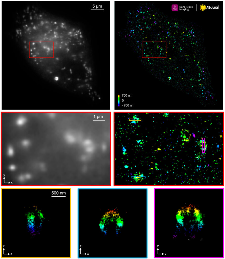
The STORM super-resolution (SR) imaging of HeLa cells using SQSTM1/p62 Rabbit mAb at dilution of 1:200 with 3% paraformaldehyde (PFA) +0.1% glutaraldehyde (GA) fixation. The immunostaining was performed by Full Automatic Immunofluorescence Workflow System (Workflow Ultra300, Nano-Micro imaging, China). Image was performed with Single-Molecule Localization Super-Resolution Microscopy (STORM Ultra300, Nano-Micro imaging.
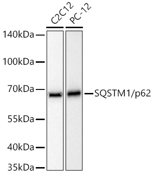
Western blot analysis of various lysates using SQSTM1/p62 Rabbit mAb at
1:20000 dilution incubated overnight at 4℃.
Secondary antibody: HRP-conjugated Goat anti-Rabbit IgG (H+L) at 1:10000
dilution.
Lysates/proteins: 25 μg per lane.
Blocking buffer: 3% nonfat dry milk in TBST.
Detection: ECL West Pico Plus.
Exposure time: 10s.
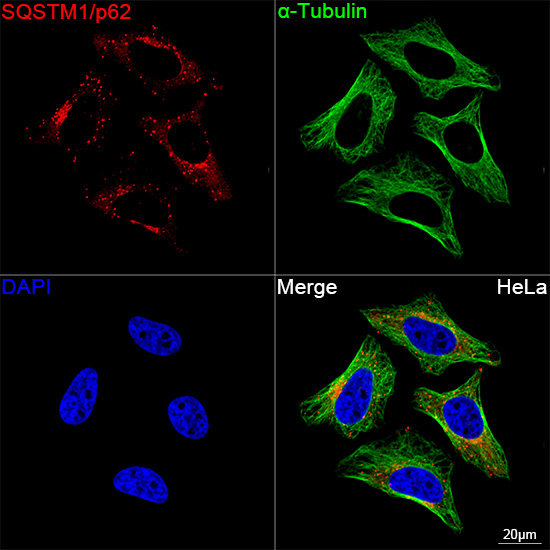
Confocal imaging of HeLa cells using SQSTM1/p62 Rabbit mAb (dilution 1:200) followed by a further incubation with Cy3 Goat Anti-Rabbit IgG (H+L) ( dilution 1:500) (Red). The cells were counterstained with α-Tubulin Mouse mAb ( dilution 1:400) followed by incubation with conjugated Goat Anti-Mouse IgG (H+L) Ab ( dilution 1:500) (Green). DAPI was used for nuclear staining (Blue). Objective: 100x.