SYN1 Mouse Monoclonal Antibody
| Cat Number: | MABN81677 |
|---|---|
| Conjugate: | Unconjugated |
| Size: | 100ul |
| Concentration: | 1mg/ml |
| Host: | Mouse |
| Isotype: | Mouse IgG1 |
| Immunogen: | Purified recombinant fragment of human SYN1 (AA: 362-511) expressed in E. Coli. |
| Reactivity: | Human,Mouse,Monkey,Rat |
| Applications: | WB 1:500-1:2000,IHC 1:200-1:1000,ICC 1:200-1:1000,ELISA 1:5000-1:20000,FC 1:200-1:400 |
| Molecular: | 1kDa |
| Purification: | Affinity purification |
| Synonyms: | SYNI; SYN1a; SYN1b |
| Background: | This gene is a member of the synapsin gene family. Synapsins encode neuronal phosphoproteins which associate with the cytoplasmic surface of synaptic vesicles. Family members are characterized by common protein domains, and they are implicated in synaptogenesis and the modulation of neurotransmitter release, suggesting a potential role in several neuropsychiatric diseases. This member of the synapsin family plays a role in regulation of axonogenesis and synaptogenesis. The protein encoded serves as a substrate for several different protein kinases and phosphorylation may function in the regulation of this protein in the nerve terminal. Mutations in this gene may be associated with X-linked disorders with primary neuronal degeneration such as Rett syndrome. Alternatively spliced transcript variants encoding different isoforms have been identified. |
| Form: | liquid |
| Buffer: | Purified antibody in PBS with 0.05% sodium azide |
| Storage: | Store at 4°C short term. Aliquot and store at -20°C long term. Avoid freeze/thaw cycles. |
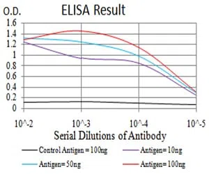
Black line: Control Antigen (100 ng);Purple line: Antigen (10ng); Blue line: Antigen (50 ng); Red line:Antigen (100 ng)
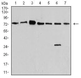
Western blot analysis using SYN1 mouse mAb against SK-N-SH (1), NIH/3T3 (2), U251 (3), C6 (4), A549 (5), MCF-7 (6), and COS7 (7) cell lysate.
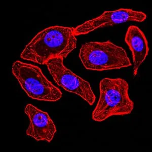
Immunofluorescence analysis of GC-7901 cells using SYN1 mouse mAb (green). Blue: DRAQ5 fluorescent DNA dye. Red: Actin filaments have been labeled with Alexa Fluor- 555 phalloidin.