
Synuclein Alpha Chicken Polyclonal Antibody
| Cat Number: | AB-83921 |
|---|---|
| Conjugate: | Unconjugated |
| Size: | 100 ug |
| Clone: | POLY |
| Concentration: | 1mg/ml |
| Host: | Chicken |
| Isotype: | IgY |
| Immunogen: | Full length recombinant human protein expressed in and purified from E. coli |
| Reactivity: | Human, rat, mouse, cow, pig, horse |
| Applications: | WB: 1:2,000. IF/ICC and IHC: 1:2,000-1:5,000. |
| Molecular: | 15kDa by SDSPAGE |
| Purification: | Purified |
| Background: | The CPCA-SNCA antibody recognizes full length human and rodent α-synuclein specifically both in western blots and in immunocytochemical experiments and is a good marker of synapses on rodents,including transgenic mice. We document that the antibody works well not only for western blotting, IF and ICC but also on formalin fixed paraffin embedded sections, select the “Additional Data” for this data. We also market a mouse monoclonal to α-synuclein. The 2A7 epitope is in the region 61-95 which corresponding to the central “non-amyloid beta component of Alzheimer’s disease amyloid”, a proteolytic fragment of α-synuclein which co-purified with Alzheimer’s amyloid. This monoclonal antibody has been used as an ELISA capture reagent capable of detecting α-synuclein in human plasma in combination with a polyclonal antibody such as SNCA. |
| Form: | Liquid |
| Buffer: | Immunogen affinity purified antibody at 1mg/mL in 50% PBS, 50% glycerol plus 5mM NaN3 |
| Storage: | Store at 4°C, for longer term store at -20°C. Stable for 12 months from date of receipt. |
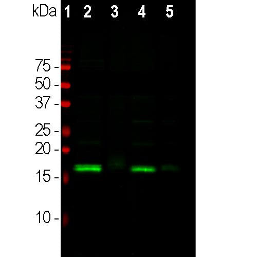
Western blot analysis of different tissue lysates using
chicken pAb to α-synuclein, CPCA-SNCA, dilution 1:2,000 in green: [1] protein standard (red), [2] rat brain, [3] rat spinal cord, [4] mouse brain, [5] mouse spinal cord. The strong band at about 15kDa corresponds to the α- synuclein protein in brain extracts, which are rich in
synapses, while a weaker band is seen in spinal cord
extracts where synapses are a more minor component.
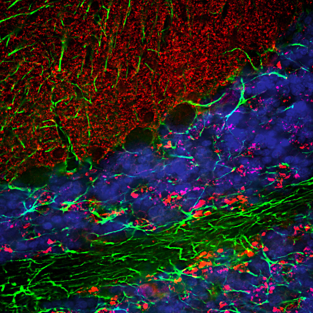
Immunofluorescent analysis of a section of rat cerebellum stained with chicken pAb to α-synuclein, dilution 1:3,000 in red, and costained with rabbit pAb to GFAP, GFAP, dilution 1:5,000 in green. The blue isDAPI staining of nuclear DNA. Following transcardial
perfusion of rat with 4% paraformaldehyde, brain was post fixed for 24 hours, cut to 45μM, and free-floating sections were stained with above antibodies. The α-synuclein protein is concentrated in presynaptic regions in the granular and molecular layers, while GFAP antibody stains the network of Bergmann and astroglial cells.