TGFβ1 Rabbit Polyclonal Antibody
| Cat Number: | ABN18858 |
|---|---|
| Conjugate: | Unconjugated |
| Size: | 100μL |
| Clone: | POLY |
| Concentration: | 1mg/ml |
| Host: | Rabbit |
| Isotype: | IgG |
| Immunogen: | The antiserum was produced against synthesized peptide derived from human TGF beta1. AA range:336-385 |
| Reactivity: | Human,Mouse,Rat |
| Applications: | WB 1:500-1:2000,IHC 1:100-1:300,ICC/IF 1:100-1:300,ELISA 1:10000-1:20000 |
| Molecular: | 55kDa |
| Purification: | Affinity purification |
| Synonyms: | TGFB1; TGFB; Transforming growth factor beta-1; TGF-beta-1 |
| Background: | This gene encodes a secreted ligand of the TGF-beta (transforming growth factor-beta) superfamily of proteins. Ligands of this family bind various TGF-beta receptors leading to recruitment and activation of SMAD family transcription factors that regulate gene expression. The encoded preproprotein is proteolytically processed to generate a latency-associated peptide (LAP) and a mature peptide, and is found in either a latent form composed of a mature peptide homodimer, a LAP homodimer, and a latent TGF-beta binding protein, or in an active form consisting solely of the mature peptide homodimer. The mature peptide may also form heterodimers with other TGFB family members. This encoded protein regulates cell proliferation, differentiation and growth, and can modulate expression and activation of other growth factors including interferon gamma and tumor necrosis factor alpha. This gene idisease:Defects in TGFB1 are the cause of Camurati-Engelmann disease (CED) [MIM:131300]; also known as progressive diaphyseal dysplasia 1 (DPD1). CED is an autosomal dominant disorder characterized by hyperostosis and sclerosis of the diaphyses of long bones. The disease typically presents in early childhood with pain, muscular weakness and waddling gait, and in some cases other features such as exophthalmos, facial paralysis, hearing difficulties and loss of vision.,function:Multifunctional protein that controls proliferation, differentiation and other functions in many cell types. Many cells synthesize TGFB1 and have specific receptors for it. It positively and negatively regulates many other growth factors. It plays an important role in bone remodeling as it is a potent stimulator of osteoblastic bone formation, causing chemotaxis, proliferation and differentiation in committed osteoblasts.,induction:Activated in vitro at pH below 3.5 and over 12.5.,online information:TGF beta-1 entry,polymorphism:In post-menopausal Japanese women, the frequency of Leu-10 is higher in subjects with osteoporosis than in controls.,PTM:Glycosylated.,PTM:The precursor is cleaved into mature TGF-beta-1 and LAP, which remains non-covalently linked to mature TGF-beta-1 rendering it inactive.,similarity:Belongs to the TGF-beta family.,subunit:The inactive form consists of a TGFB1 homodimer non-covalently linked to a latency-associated peptide (LAP) homodimer. The inactive complex can contain a latent TGFB1-binding protein. The active form is a homodimer of mature TGFB1; disulfide-linked. Heterodimers of TGFB1/TGFB2 have been found in bone. Interacts with CD109 and DPT.,tissue specificity:Highly expressed in bone., |
| Form: | liquid |
| Buffer: | Liquid in PBS containing 50% glycerol, 0.5% protective protein and 0.02% New type preservative N. |
| Storage: | Store at 4°C short term. Aliquot and store at -20°C long term. Avoid freeze/thaw cycles. |
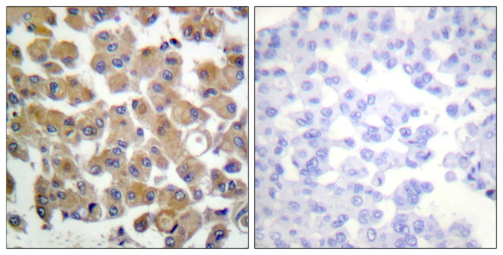
Immunohistochemistry analysis of paraffin-embedded human breast carcinoma
tissue, using TGF beta1 Antibody. The picture on the right is blocked with the
synthesized peptide.
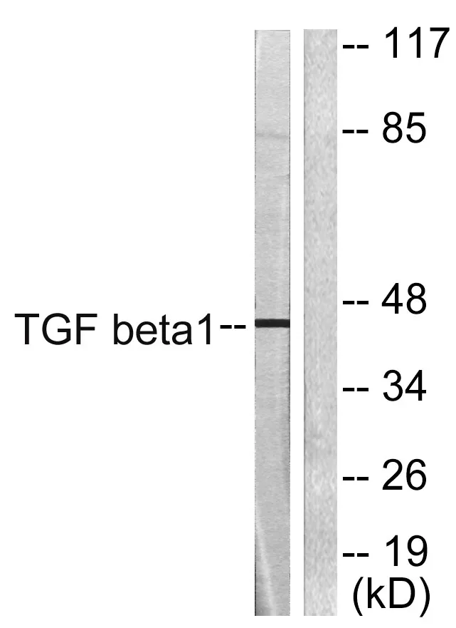
Western blot analysis of lysates from HepG2 cells, using TGF beta1 Antibody. The
lane on the right is blocked with the synthesized peptide.

Immunofluorescence analysis of rat-lung tissue. 1,TGFβ1 Polyclonal Antibody(red) was diluted at 1:200(4°C,overnight). 2, Cy3 labled Secondary antibody was diluted at 1:300(room temperature, 50min).3, Picture B: DAPI(blue) 10min. Picture A:Target. Picture B: DAPI. Picture C: merge of A+B