TNF-α Rabbit Polyclonal Antibody
| Cat Number: | ABE3623 |
|---|---|
| Conjugate: | Unconjugated |
| Size: | 100 ug |
| Concentration: | 1mg/ml |
| Host: | Rabbit |
| Isotype: | IgG |
| Immunogen: | Recombinant protein.This information is considered to be commercially sensitive. |
| Reactivity: | Human,Mouse,Rat |
| Applications: | WB 1:500 - 1:2000 IHC-P 1:500 - 1:1000 IF/ICC 1:50 - 1:100 ELISA Recommended starting concentration is 1 μg/mL. Please optimize the concentration based on your specific assay requirements. |
| Molecular: | 18kDa/25kDa |
| Purification: | Affinity purification |
| Synonyms: | DIF; TNFA; TNFSF2; TNLG1F; TNF-alpha; TNF-α |
| Background: | This gene encodes a multifunctional proinflammatory cytokine that belongs to the tumor necrosis factor (TNF) superfamily. This cytokine is mainly secreted by macrophages. It can bind to, and thus functions through its receptors TNFRSF1A/TNFR1 and TNFRSF1B/TNFBR. This cytokine is involved in the regulation of a wide spectrum of biological processes including cell proliferation, differentiation, apoptosis, lipid metabolism, and coagulation. This cytokine has been implicated in a variety of diseases, including autoimmune diseases, insulin resistance, psoriasis, rheumatoid arthritis ankylosing spondylitis, tuberculosis, autosomal dominant polycystic kidney disease, and cancer. Mutations in this gene affect susceptibility to cerebral malaria, septic shock, and Alzheimer disease. Knockout studies in mice also suggested the neuroprotective function of this cytokine. |
| Form: | liquid |
| Buffer: | PBS with 0.09% Sodium azide,50% glycerol,pH7.3. |
| Storage: | Store at -20℃. Avoid freeze / thaw cycles. |
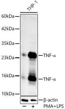
Western blot analysis of lysates from THP-1 cells, using TNF-α Rabbit pAb at 1:1000 dilution. THP-1 cells were treated with PMA/TPA (80 nM) at 37℃ for overnight and LPS (1 μg/ml) at 37℃ for 6 hours.
Secondary antibody: HRP-conjugated Goat anti-Rabbit IgG (H+L) at 1:10000 dilution.
Lysates/proteins: 25μg per lane.
Blocking buffer: 3% nonfat dry milk in TBST.
Detection: ECL West Pico Plus.
Exposure time: 60s.
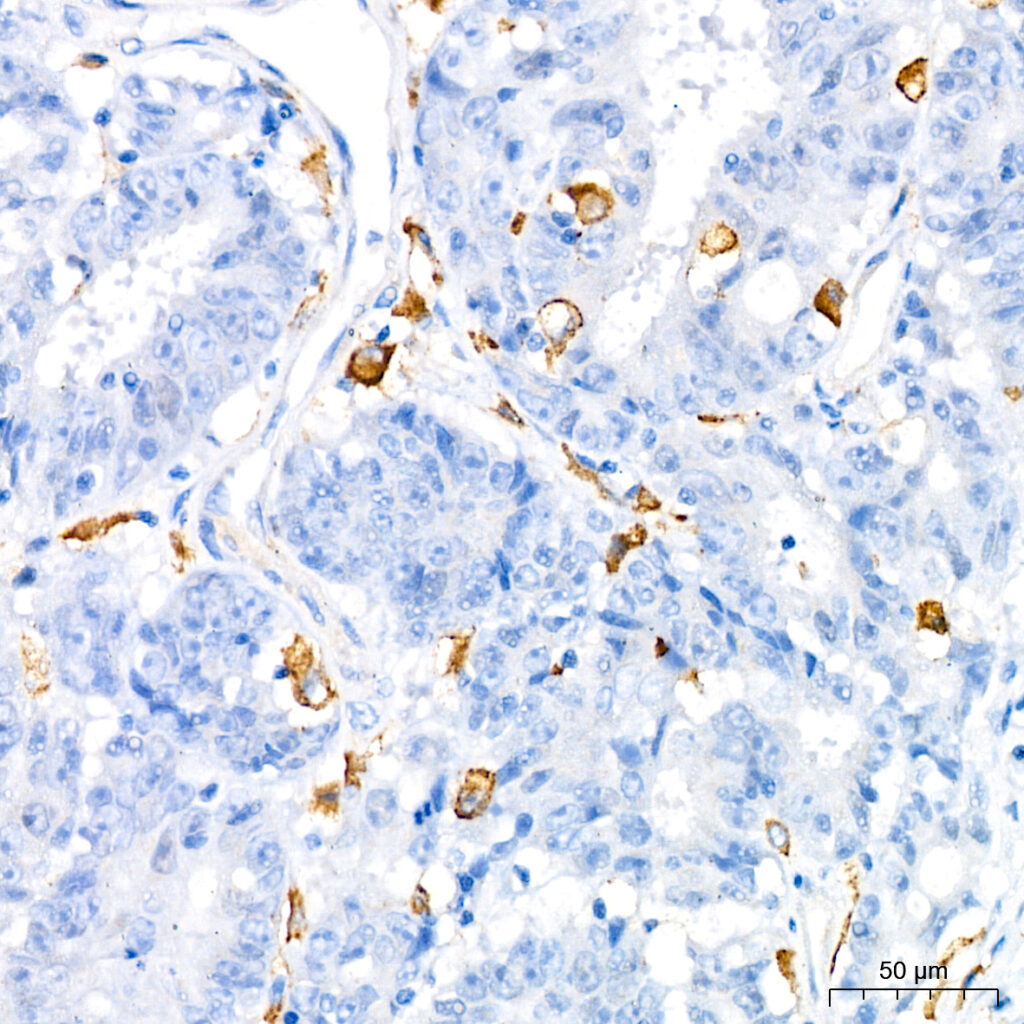
Immunohistochemistry analysis of paraffin-embedded Human colon carcinoma using TNF-α Rabbit pAb ) at dilution of 1:1000 (40x lens). High pressure antigen retrieval performed with 0.01M Citrate buffer (pH 6.0) prior to IHC staining.
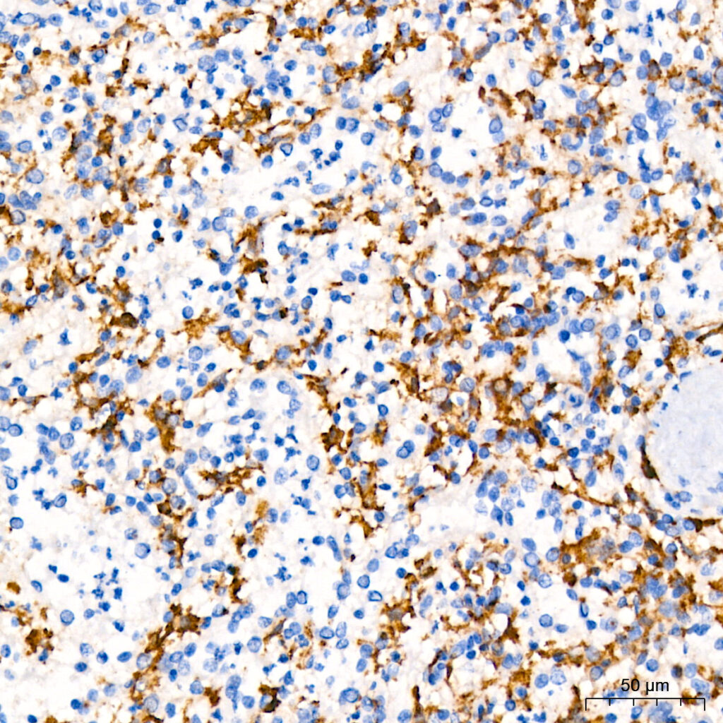
Immunohistochemistry analysis of paraffin-embedded Human spleen using TNF-α Rabbit pAb ) at dilution of 1:1000 (40x lens). High pressure antigen retrieval performed with 0.01M Citrate buffer (pH 6.0) prior to IHC staining.