Tsg101 rabbit Polyclonal Antibody
| Cat Number: | ABE7453 |
|---|---|
| Conjugate: | unconjugated |
| Size: | 100 ug |
| Clone: | Polyclonal |
| Concentration: | 1 mg/ml |
| Host: | Rabbit |
| Isotype: | IgG |
| Immunogen: | The antiserum was produced against synthesized peptide derived from human TSG101. AA range:281-330 |
| Reactivity: | Human;Mouse;Rat;Monkey |
| Applications: | Western Blot: 1/500 - 1/2000. Immunohistochemistry: 1/100 - 1/300. Immunofluorescence: 1/200 - 1/1000. ELISA: 1/5000. Not yet tested in other applications. |
| Molecular: | 44kD |
| Purification: | The antibody was affinity-purified fromrabbit antiserum by affinity-chromatography using epitope-specific immunogen. |
| Synonyms: | TSG101; Tumor susceptibility gene 101 protein; ESCRT-I complex subunit TSG101 |
| Background: | The protein encoded by this gene belongs to a group of apparently inactive homologs of ubiquitin-conjugating enzymes. The gene product contains a coiled-coil domain that interacts with stathmin, a cytosolic phosphoprotein implicated in tumorigenesis. The protein may play a role in cell growth and differentiation and act as a negative growth regulator. In vitro steady-state expression of this tumor susceptibility gene appears to be important for maintenance of genomic stability and cell cycle regulation. Mutations and alternative splicing in this gene occur in high frequency in breast cancer and suggest that defects occur during breast cancer tumorigenesis and/or progression. [provided by RefSeq, Jul 2008], |
| Form: | liquid |
| Buffer: | Liquid in PBS containing 50% glycerol, 0.5%BSAand0.02% sodium azide. |
| Storage: | -20°C/1 year |

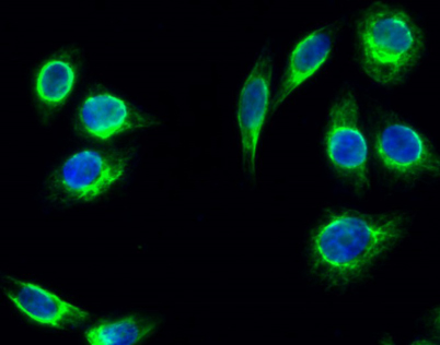
Immunofluorescence analysis of Hela cell. 1,Tsg 101 Polyclonal Antibody(green) was diluted at 1:200(4° overnight). 2, Goat Anti Rabbit Alexa Fluor 488 was diluted at 1:1000(room temperature, 50min). 3 DAPI(blue) 10min.

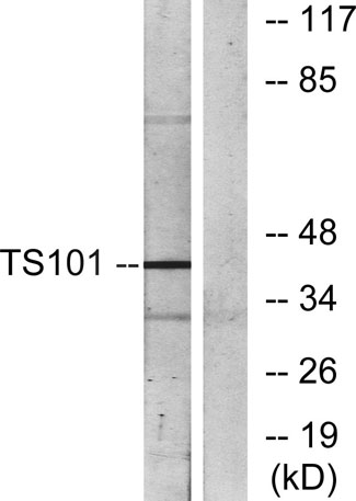
Western blot analysis of lysates from COS7 cells, using TS101 Antibody. The lane on the right is blocked with the synthesized peptide.

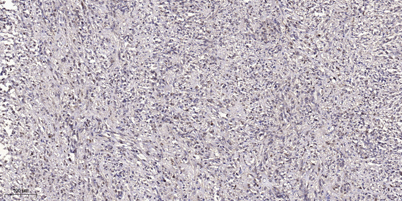
Immunohistochemical analysis of paraffin-embedded human small intestinal carcinoma tissue. 1,primary Antibody was diluted at 1:200(4° overnight). 2, Sodium citrate pH 6.0 was used for antigen retrieval(>98°C,20min). 3,Secondary antibody was diluted at 1:2
