TTR Rabbit Polyclonal Antibody
| Cat Number: | AB1120 |
|---|---|
| Conjugate: | Unconjugated |
| Size: | 100 ug |
| Concentration: | 1mg/ml |
| Host: | Rabbit |
| Isotype: | IgG |
| Immunogen: | Recombinant protein.This information is considered to be commercially sensitive. |
| Reactivity: | Human, Mouse, Rat |
| Applications: | WB 1:500 - 1:1000 IHC-P 1:50 - 1:100 IF/ICC 1:50 - 1:100 ELISA Recommended starting concentration is 1 μg/mL. Please optimize the concentration based on your specific assay requirements. |
| Purification: | Affinity purification |
| Synonyms: | CTS; TTN; ATTR; CTS1; PALB; TBPA; HEL111; HsT2651; TTR |
| Background: | This gene encodes one of the three prealbumins, which include alpha-1-antitrypsin, transthyretin and orosomucoid. The encoded protein, transthyretin, is a homo-tetrameric carrier protein, which transports thyroid hormones in the plasma and cerebrospinal fluid. It is also involved in the transport of retinol (vitamin A) in the plasma by associating with retinol-binding protein. The protein may also be involved in other intracellular processes including proteolysis, nerve regeneration, autophagy and glucose homeostasis. Mutations in this gene are associated with amyloid deposition, predominantly affecting peripheral nerves or the heart, while a small percentage of the gene mutations are non-amyloidogenic. The mutations are implicated in the etiology of several diseases, including amyloidotic polyneuropathy, euthyroid hyperthyroxinaemia, amyloidotic vitreous opacities, cardiomyopathy, oculoleptomeningeal amyloidosis, meningocerebrovascular amyloidosis and carpal tunnel syndrome. |
| Form: | Liquid |
| Buffer: | PBS with 0.09% Sodium azide, 50% glycerol, pH7.3. |
| Storage: | Store at -20℃. Avoid freeze / thaw cycles. |

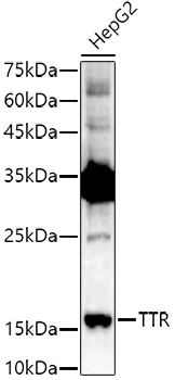
Western blot analysis of lysates from HepG2 cells, using TTR Rabbit Polyclonal Antibody at 1:1000 dilution.
Secondary antibody: HRP-conjugated Goat anti-Rabbit IgG (H+L) at 1:10000 dilution.
Lysates/proteins: 25μg per lane.
Blocking buffer: 3% nonfat dry milk in TBST.
Detection: ECL Enhanced Kit.
Exposure time: 180s.

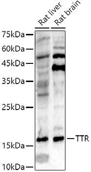
Western blot analysis of various lysates using TTR Rabbit Polyclonal Antibody at 1:500 dilution.
Secondary antibody: HRP-conjugated Goat anti-Rabbit IgG (H+L) at 1:10000 dilution.
Lysates/proteins: 25 μg per lane.
Blocking buffer: 3% nonfat dry milk in TBST.
Detection: ECL Enhanced Kit.
Exposure time: 180s.

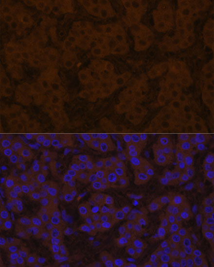
Immunofluorescence analysis of human liver cancer using TTR Rabbit Polyclonal Antibody at dilution of 1:100 (40x lens). Blue: DAPI for nuclear staining.
