
Vitronectin Rabbit pAb
| Cat Number: | AB1667 |
|---|---|
| Conjugate: | Unconjugated |
| Size: | 100 ug |
| Host: | Rabbit |
| Isotype: | IgG |
| Immunogen: | Recombinant protein (or fragment).This information is considered to be commercially sensitive. |
| Reactivity: | Human,Mouse,Rat |
| Applications: | WB 1:500 - 1:2000; IHC-P 1:50 - 1:200; ELISA Recommended starting concentration is 1 μg/mL. Please optimize the concentration based on your specific assay requirements. |
| Molecular: | Observed MW: 65kDa, 75kDa; Calculated MW: 54kDa |
| Purification: | Affinity purification |
| Synonyms: | VN; V75; VNT; Vitronectin |
| Background: | The protein encoded by this gene functions in part as an adhesive glycoprotein. Differential expression of this protein can promote either cell adhesion or migration as it links cells to the extracellular matrix through a variety of ligands. These ligands include integrins, plasminogen activator inhibitor-1, and urokinase plasminogen activator receptor. This secreted protein can be present in the plasma as a monomer or dimer and forms a multimer in the extracellular matrix of several tissues. This protein also inhibits the membrane-damaging effect of the terminal cytolytic complement pathway and binds to several serpin serine protease inhibitors. This protein can also promote extracellular matrix degradation and thus plays a role in tumorigenesis. It is involved in a variety of other biological processes such as the regulation of the coagulation pathway, wound healing, and tissue remodeling. The heparin-binding domain of this protein give it anti-microbial properties. It is also a lipid binding protein that forms a principal component of high density lipoprotein. |
| Form: | Liquid |
| Buffer: | PBS containing 50% glycerol, preserved with proclin300 or sodium azide (as specified on the Certificate of Analysis), pH 7.3. |
| Storage: | Store at -20℃. Avoid freeze / thaw cycles. |
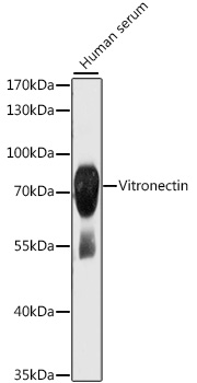
Western blot analysis of lysates from human serum, using Vitronectin Rabbit pAb ) at 1:1000 dilution. Secondary antibody: HRP-conjugated Goat anti-Rabbit IgG (H+L) at 1:10000 dilution. Lysates/proteins: 25μg per lane. Blocking buffer: 3% nonfat dry milk in TBST. Detection: ECL Basic Kit.
Exposure time: 1s.
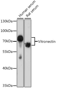
Western blot analysis of various lysates using Vitronectin Rabbit pAb at 1:1000 dilution. Secondary antibody: HRP-conjugated Goat anti-Rabbit IgG (H+L) at 1:10000 dilution. Lysates/proteins: 25μg per lane. Blocking buffer: 3% nonfat dry milk in TBST. Detection: ECL Basic Kit.
Exposure time: 20s.
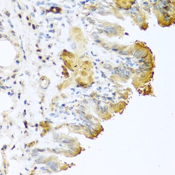
Immunohistochemistry analysis of paraffin-embedded Mouse lung using Vitronectin Rabbit pAb at dilution of 1:100 (40x lens). Microwave antigen retrieval performed with 0.01M PBS Buffer (pH 7.2) prior to IHC staining.