XRCC1 Rabbit Polyclonal Antibody
| Cat Number: | ABN19971 |
|---|---|
| Conjugate: | Unconjugated |
| Size: | 100μL |
| Clone: | POLY |
| Concentration: | 1mg/ml |
| Host: | Rabbit |
| Isotype: | IgG |
| Immunogen: | The antiserum was produced against synthesized peptide derived from human XRCC1. AA range:517-566. |
| Reactivity: | Human,Mouse,Rat |
| Applications: | WB 1:500-1:2000,IHC 1:100-1:300,ICC/IF 1:50-1:200,ELISA 1:5000-1:20000 |
| Molecular: | 70kDa |
| Purification: | Affinity purification |
| Synonyms: | XRCC1; DNA repair protein XRCC1; X-ray repair cross-complementing protein 1 |
| Background: | The protein encoded by this gene is involved in the efficient repair of DNA single-strand breaks formed by exposure to ionizing radiation and alkylating agents. This protein interacts with DNA ligase III, polymerase beta and poly (ADP-ribose) polymerase to participate in the base excision repair pathway. It may play a role in DNA processing during meiogenesis and recombination in germ cells. A rare microsatellite polymorphism in this gene is associated with cancer in patients of varying radiosensitivity. [provided by RefSeq, Jul 2008],function:Corrects defective DNA strand-break repair and sister chromatid exchange following treatment with ionizing radiation and alkylating agents.,polymorphism:Carriers of the polymorphic Gln-399 allele may be at greater risk for tobacco- and age-related DNA damage.,PTM:Phosphorylation of Ser-371 causes dimer dissociation. Phosphorylation by CK2 promotes interaction with APTX and APLF.,PTM:Sumoylated.,similarity:Contains 2 BRCT domains.,subcellular location:Accumulates at sites of DNA damage.,subunit:Homodimer. Interacts with polynucleotide kinase (PNK), DNA polymerase-beta (POLB) and DNA ligase III (LIG3). Interacts with APTX and APLF., |
| Form: | liquid |
| Buffer: | Liquid in PBS containing 50% glycerol, 0.5% protective protein and 0.02% New type preservative N. |
| Storage: | Store at 4°C short term. Aliquot and store at -20°C long term. Avoid freeze/thaw cycles. |
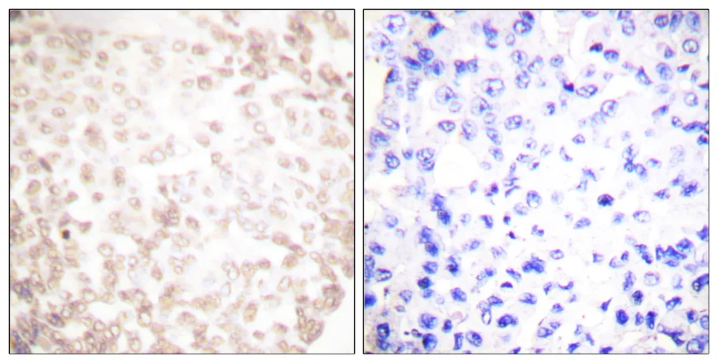
Immunohistochemistry analysis of paraffin-embedded human lung carcinoma tissue, using XRCC1 Antibody. The picture on the right is blocked with the synthesized peptide.
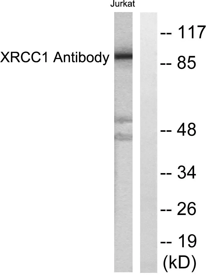
Western blot analysis of lysates from Jurkat cells, using XRCC1 Antibody. The lane on the right is blocked with the synthesized peptide.
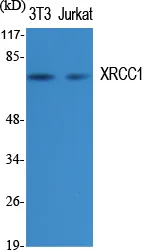
Western Blot analysis of various cells using XRCC1 Polyclonal Antibody diluted at 1:1000. Secondary antibody was diluted at 1:20000.