β-Tubulin Rabbit Monoclonal Antibody
| Cat Number: | MAB12289 |
|---|---|
| Conjugate: | Unconjugated |
| Size: | 100 ug |
| Clone: | 9F3 |
| Concentration: | 1mg/ml |
| Host: | Rabbit |
| Isotype: | IgG |
| Immunogen: | A synthetic peptide corresponding to a sequence within amino acids 250-350 of human β-Tubulin (P07437). |
| Reactivity: | Human,Mouse,Rat |
| Applications: | WB,1:5000 - 1:10000 IHC-P,1:50 - 1:200 IP,0.5μg-4μg antibody for 200μg-400μg extracts of whole cells ELISA,Recommended starting concentration is 1 μg/mL. Please optimize the concentration based on your specific assay requirements. |
| Molecular: | 55kDa |
| Purification: | Affinity purification |
| Background: | This gene encodes a beta tubulin protein. This protein forms a dimer with alpha tubulin and acts as a structural component of microtubules. Mutations in this gene cause cortical dysplasia, complex, with other brain malformations 6. Alternative splicing results in multiple splice variants. There are multiple pseudogenes for this gene on chromosomes 1, 6, 7, 8, 9, and 13. |
| Form: | liquid |
| Buffer: | PBS with 0.02% Sodium azide,0.05% BSA,50% glycerol,pH7.3. |
| Storage: | Store at -20℃. Avoid freeze / thaw cycles. |

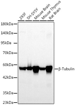
Western blot analysis of various lysates using β-Tubulin Rabbit mAb at 1:5000 dilution.
Secondary antibody: HRP-conjugated Goat anti-Rabbit IgG at 1:10000 dilution.
Lysates/proteins: 25μg per lane.
Blocking buffer: 3% nonfat dry milk in TBST.
Detection: ECL Basic Kit .
Exposure time: 20s.

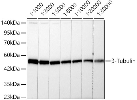
Western blot analysis of lysates from 293F cells, using β-Tubulin Rabbit mAb at 1:1000-1:30000 dilution.
Secondary antibody: HRP-conjugated Goat anti-Rabbit IgG at 1:10000 dilution.
Lysates/proteins: 25μg per lane.
Blocking buffer: 3% nonfat dry milk in TBST.
Detection: ECL Basic Kit .
Exposure time: 10s.

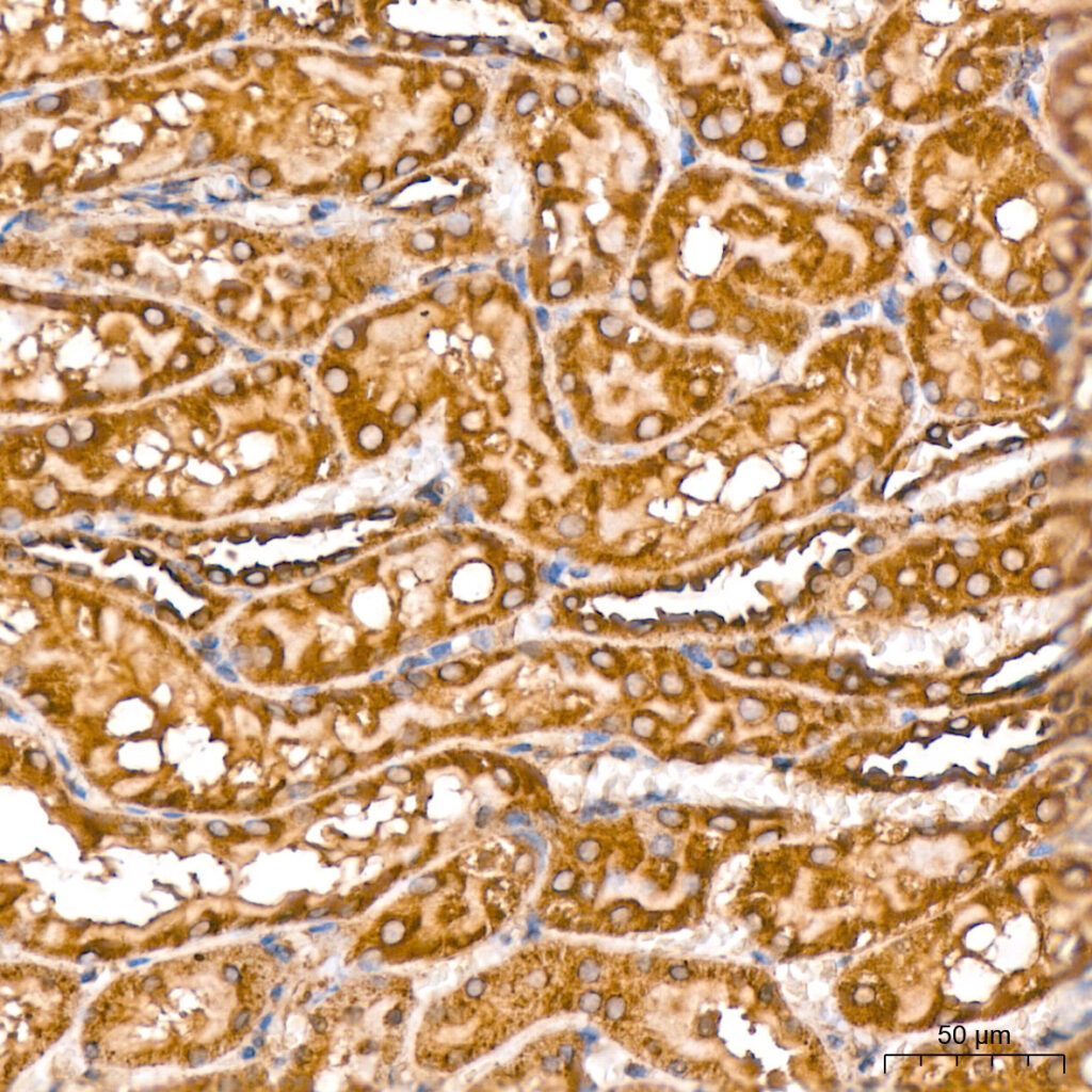
Immunohistochemistry analysis of paraffin-embedded Rat kidney tissue using β-Tubulin Rabbit mAb at a dilution of 1:200 . High pressure antigen retrieval performed with 0.01M Citrate Bufferr prior to IHC staining.
