
MCL-1 Mouse Monoclonal Antibody
| Cat Number: | MABN80516 |
|---|---|
| Conjugate: | Unconjugated |
| Size: | 100μL |
| Concentration: | 1mg/ml |
| Host: | Mouse |
| Isotype: | Mouse IgG1 |
| Reactivity: | Human |
| Applications: | WB 1:500-1:2000,IHC 1:200-1:1000,ICC 1:200-1:1000,ELISA 1:5000-1:20000 |
| Molecular: | 37kDa |
| Purification: | Affinity purification |
| Synonyms: | EAT, MCL1L, MCL1S |
| Background: | Mcl-1 (Myeloid cell leukemia-1) is Bcl-2-related and was identified as an early-induction gene thatincreased in expression during the differentiation of human myeloblastic leukemia cell ML-1, orexposure to different DNA damaging agents. The level of Mcl-1 is decreased in peripheral B lymphocytes undergoing apoptosis following treatment with apoptotic stimuli such as TGF-alpha 1 and forskolin. Expression of Mcl-1 is able to delay apoptosis induced by over-expression of c-myc in CHO 5AHSmyc cells. In hematopoietic FDC-P1 cells, Mcl-1 interacts with another Bcl-2-relatedprotein, Bax, and prolongs cell viability after treatment with different apoptotic reagents.Thismonoclonal antibody detected a 37kd MCL1 in BCBL-1 cell lysate. |
| Form: | liquid |
| Buffer: | Purified antibody in PBS with 0.05% sodium azide |
| Storage: | Store at 4°C short term. Aliquot and store at -20°C long term. Avoid freeze/thaw cycles. |
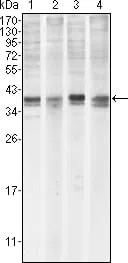
Western blot analysis using MCL1 mouse mAb against Hela (1), BCBL-1 (2), Jurkat (3) and HL60 (4) cell lysate.
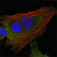
Confocal Immunofluorescence analysis of HepG2 cells using MCL1 mouse mAb (green). Red: Actin filaments have been labeled with DY-554 phalloidin. Blue: DRAQ5 fluorescent DNA dye.
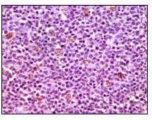
Immunohistochemical analysis of paraffin-embedded human lymphnode tissues using MCL1 mouse mAb with DAB staining.