
p21 rabbit pAb
| Cat Number: | ABE3100 |
|---|---|
| Conjugate: | unconjugated |
| Size: | 100 ug |
| Clone: | Polyclonal |
| Concentration: | 1 mg/ml |
| Host: | Rabbit |
| Isotype: | IgG |
| Immunogen: | The antiserum was produced against synthesized peptide derived from human p21 Cip1. AA range:111-160 |
| Reactivity: | Human;Mouse;Rat |
| Applications: | IF: 1:50-200 Western Blot: 1/500 - 1/2000. ELISA: 1/20000. Not yet tested in other applications. |
| Molecular: | 21kD |
| Synonyms: | CDKN1A; CAP20; CDKN1; CIP1; MDA6; PIC1; SDI1; WAF1; Cyclin-dependent kinase inhibitor 1; CDK-interacting protein 1; Melanoma differentiation-associated protein 6; MDA-6; p21 |
| Background: | This gene encodes a potent cyclin-dependent kinase inhibitor. The encoded protein binds to and inhibits the activity of cyclin-cyclin-dependent kinase2 or -cyclin-dependent kinase4 complexes, and thus functions as a regulator of cell cycle progression at G1. The expression of this gene is tightly controlled by the tumor suppressor protein p53, through which this protein mediates the p53-dependent cell cycle G1 phase arrest in response to a variety of stress stimuli. This protein can interact with proliferating cell nuclear antigen, a DNA polymerase accessory factor, and plays a regulatory role in S phase DNA replication and DNA damage repair. This protein was reported to be specifically cleaved by CASP3-like caspases, which thus leads to a dramatic activation of cyclin-dependent kinase2, and may be instrumental in the execution of apoptosis following caspase activation. Mice that lac |
| Form: | liquid |
| Storage: | -20°C/1 year |
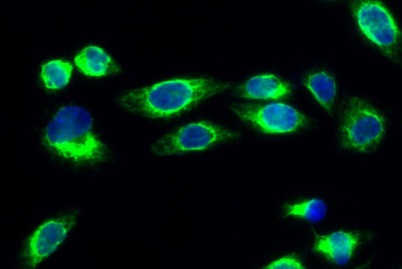
Immunofluorescence analysis of Hela cell. 1,p21 Polyclonal Antibody(green) was diluted at 1:200(4° overnight). 2, Goat Anti Rabbit Alexa Fluor 488 was diluted at 1:1000(room temperature, 50min).
3 DAPI(blue) 10min.
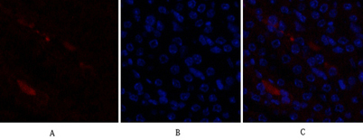
Immunofluorescence analysis of mouse-kidney tissue.
1,p21 Polyclonal Antibody(red) was diluted at 1:200(4°C,overnight).
2, Cy3 labled Secondary antibody was diluted at 1:300(room temperature, 50min).
3, Picture B: DAPI(blue) 10min. Picture A:Target. Pictur
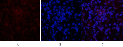
Immunofluorescence analysis of rat-lung tissue.
1,p21 Polyclonal Antibody(red) was diluted at 1:200(4°C,overnight).
2, Cy3 labled Secondary antibody was diluted at 1:300(room temperature, 50min).
3, Picture B: DAPI(blue) 10min. Picture A:Target. Picture B: