Sp1 Rabbit Polyclonal Antibody
| Cat Number: | ABE3482 |
|---|---|
| Conjugate: | Unconjugated |
| Size: | 100 ug |
| Clone: | Polyclonal |
| Concentration: | 1 mg/ml |
| Host: | Rabbit |
| Isotype: | IgG |
| Immunogen: | The antiserum was produced against synthesized peptide derived from human SP1. AA range:706-755 |
| Reactivity: | Human;Mouse;Rat |
| Applications: | Western Blot: 1/500 - 1/2000. Immunohistochemistry: 1/100 - 1/300. Immunofluorescence: 1/200 - 1/1000. ELISA: 1/20000. Not yet tested in other applications. |
| Molecular: | 85kD |
| Purification: | The antibody was affinity-purified fromrabbit antiserum by affinity-chromatography using epitope-specific immunogen. |
| Synonyms: | SP1; TSFP1; Transcription factor Sp1 |
| Background: | The protein encoded by this gene is a zinc finger transcription factor that binds to GC-rich motifs of many promoters. The encoded protein is involved in many cellular processes, including cell differentiation, cell growth, apoptosis, immune responses, response to DNA damage, and chromatin remodeling. Post-translational modifications such as phosphorylation, acetylation, glycosylation, and proteolytic processing significantly affect the activity of this protein, which can be an activator or a repressor. Three transcript variants encoding different isoforms have been found for this gene. [provided by RefSeq, Nov 2014], |
| Form: | liquid |
| Buffer: | Liquid in PBS containing 50% glycerol, 0.5%BSAand0.02% sodium azide. |
| Storage: | -20°C/1 year |
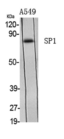
Western Blot analysis of various cells using Sp1 Polyclonal Antibody
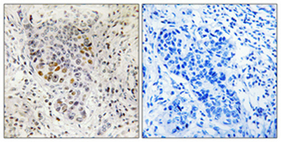
Immunohistochemical analysis of paraffin-embedded Human lung cancer. Antibody was diluted at 1:100(4° overnight). High-pressure and temperature Tris-EDTA,pH8.0 was used for antigen retrieval. Negetive contrl (right) obtaned from antibody was pre-absorbe

Immunofluorescence analysis of HepG2 cells, using SP1 Antibody. The picture on the right is blocked with the synthesized peptide.