TAK1 Rabbit Monoclonal Antibody
| Cat Number: | MABN21459 |
|---|---|
| Conjugate: | Unconjugated |
| Size: | 100 ug |
| Concentration: | 1 mg/ml. The concentration of this product may be batch-dependent. |
| Host: | Rabbit |
| Isotype: | IgG,Kappa |
| Immunogen: | A synthetic peptide corresponding to target protein. |
| Reactivity: | Human,Mouse,Rat |
| Applications: | Western Blot 1:1000-1:5000; Immunocytochemistry/Immunofluorescence: 1:200-1:1000; Immunoprecipitation:1:50-1:200; ELISA 1:5000-1:20000; |
| Molecular: | Observed MW: 67kD |
| Purification: | Protein A |
| Synonyms: | MAP3K7; TAK1; Mitogen-activated protein kinase kinase kinase 7; Transforming growth factor-beta-activated kinase 1; TGF-beta-activated kinase 1 |
| Background: | Cell localization: Cytoplasm, Membrane. The protein encoded by this gene is a member of the serine/threonine protein kinase family. This kinase mediates the signaling transduction induced by TGF beta and morphogenetic protein (BMP), and controls a variety of cell functions including transcription regulation and apoptosis. In response to IL-1, this protein forms a kinase complex including TRAF6, MAP3K7P1/TAB1 and MAP3K7P2/TAB2; this complex is required for the activation of nuclear factor kappa B. This kinase can also activate MAPK8/JNK, MAP2K4/MKK4, and thus plays a role in the cell response to environmental stresses. Four alternatively spliced transcript variants encoding distinct isoforms have been reported. |
| Form: | Liquid |
| Buffer: | PBS, 50% glycerol, 0.05% Proclin 300, 0.05% protective protein |
| Storage: | Aliquot and store at -20°C (valid for 12 months). Avoid freeze/thaw cycles. |
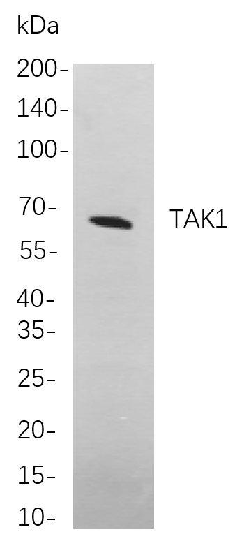
Western blot analysis of lysates from A431 cells, using TAK1 Rabbit mAb. The HRP-conjugated Goat anti-Rabbit IgG antibody was used to detect the antibody.
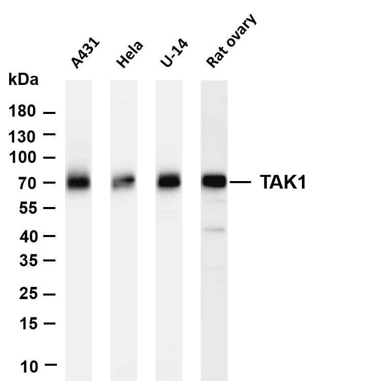
Various whole cell lysates were separated by 4-20% SDS-PAGE, and the membrane was blotted with anti-TAK1 antibody. The HRP-conjugated Goat anti-Rabbit IgG (H+L) antibody was used to detect the antibody.