Phospho-FAK (Tyr397) Rabbit Polyclonal Antibody
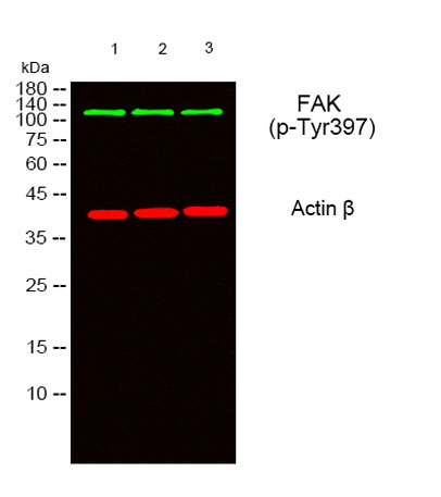
Western blot analysis of lysates from 1) 293T, 2) AD293 ,3)Hela cells, (Green) primary antibody was diluted at 1:1000, 4°over night, secondary antibody was diluted at 1:10000, 37° 1hour. (Red) Actin β Monoclonal Antibody(5B7) 
ant.

Western blot analysis of lysates from 1) 293T, 2) AD293 ,3)Hela cells, (Green) primary antibody was diluted at 1:1000, 4°over night, secondary antibody was diluted at 1:10000, 37° 1hour. (Red) Actin β Monoclonal Antibody(5B7) ant.

Catalog Number:ABP10415
Conjugate:Unconjugated
Size:100 ug
Concentration:1 mg/ml
Host:Rabbit
Isotype:IgG
Clone:POLY
Immunogen:The antiserum was produced against synthesized peptide derived from human FAK around the phosphorylation site of Tyr397. AA range:363-412.
Reactivity:Human, Rat, Mouse
Applications:Western Blot: 1:500 - 1:2000 Immunohistochemistry: 1:100 - 1:300 Immunofluorescence: 1:50 - 1:200 ELISA: 1:5000.
Molecular Weight:119kDa
Purification:The antibody was affinity-purified from rabbit antiserum by affinity-chromatography using epitope-specific immunogen.
Form:liquid
Synonyms:PTK2; FAK; FAK1; Focal adhesion kinase 1; FADK 1; Focal adhesion kinase-related nonkinase; FRNK; Protein phosphatase 1 regulatory subunit 71; PPP1R71; Protein-tyrosine kinase 2; p125FAK; pp125FAK

Applications

Western Blot: 1:500 - 1:2000 Immunohistochemistry: 1:100 - 1:300 Immunofluorescence: 1:50 - 1:200 ELISA: 1:5000.

Immunogen

The antiserum was produced against synthesized peptide derived from human FAK around the phosphorylation site of Tyr397. AA range:363-412.

Target Background

Protein tyrosine kinase 2(PTK2) Homo sapiens This gene encodes a cytoplasmic protein tyrosine kinase which is found concentrated in the focal adhesions that form between cells growing in the presence of extracellular matrix constituents. The encoded protein is a member of the FAK subfamily of protein tyrosine kinases but lacks significant sequence similarity to kinases from other subfamilies. Activation of this gene may be an important early step in cell growth and intracellular signal transduction pathways triggered in response to certain neural peptides or to cell interactions with the extracellular matrix. Several transcript variants encoding different isoforms have been found for this gene, but the full-length natures of only four of them have been determined.

Synonyms:PTK2; FAK; FAK1; Focal adhesion kinase 1; FADK 1; Focal adhesion kinase-related nonkinase; FRNK; Protein phosphatase 1 regulatory subunit 71; PPP1R71; Protein-tyrosine kinase 2; p125FAK; pp125FAK

Storage

Store at -20°C. Avoid repeated freeze-thaw cycles.

Buffer

Liquid in PBS containing 50% glycerol, 0.5% BSA and 0.02% sodium azide.

Western blot analysis of lysates from 1) 293T, 2) AD293 ,3)Hela cells, (Green) primary antibody was diluted at 1:1000, 4°over night, secondary antibody was diluted at 1:10000, 37° 1hour. (Red) Actin β Monoclonal Antibody(5B7) ant.

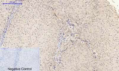
Immunohistochemical analysis of paraffin-embedded Human-liver tissue. 1,FAK (phospho Tyr397) Polyclonal Antibody was diluted at 1:200(4°C,overnight). 2, Sodium citrate pH 6.0 was used for antibody retrieval(>98°C,20min). 3,Secondary antibody was diluted at 1:200(room tempeRature, 30min). Negative control was used by secondary antibody only.

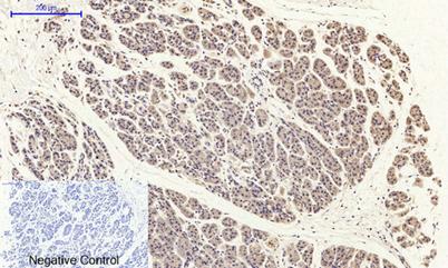
Immunohistochemical analysis of paraffin-embedded Human-stomach-cancer tissue. 1,FAK (phospho Tyr397) Polyclonal Antibody was diluted at 1:200(4°C,overnight). 2, Sodium citrate pH 6.0 was used for antibody retrieval(>98°C,20min).

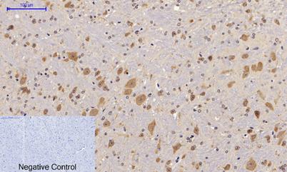
Immunohistochemical analysis of paraffin-embedded Mouse-brain tissue. 1,FAK (phospho Tyr397) Polyclonal Antibody was diluted at 1:200(4°C,overnight). 2, Sodium citrate pH 6.0 was used for antibody retrieval(>98°C,20min). 3,Secondary antibody was diluted at 1:200(room tempeRature, 30min). Negative control was used by secondary antibody only.

No documents available.