
ACE2

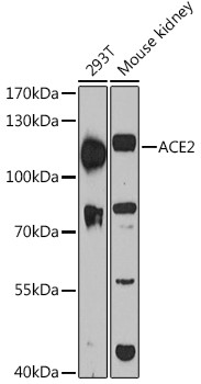
Western blot analysis of extracts of various cell lines, using ACE2 antibody at 1:3000  dilution.
Secondary antibody: HRP Goat Anti-Rabbit IgG (H+L)  at 1:10000 dilution.
Lysates/proteins: 25ug per lane.
Blocking buffer: 3% nonfat dry milk in TBST.
Detection: ECL West Pico Plus.   
Exposure time: 60s.

Western blot analysis of extracts of various cell lines, using ACE2 antibody at 1:3000 dilution. Secondary antibody: HRP Goat Anti-Rabbit IgG (H+L) at 1:10000 dilution. Lysates/proteins: 25ug per lane. Blocking buffer: 3% nonfat dry milk in TBST. Detection: ECL West Pico Plus. Exposure time: 60s.

Catalog Number:AB-84323
Size:100 ug
Concentration:1mg/ml
Host:Rb
Isotype:IgG
Clone:POLY
Immunogen:Recombinant fusion protein containing a sequence corresponding to amino acids 18-250 of human ACE2.
Reactivity:Hu, Ms, Rt
Applications:Western Blot: 1:500 - 1:2000 Immunofluorescence: 1:50 - 1:200
Molecular Weight:110kDa
Purification:Aff. Pur.
Form:Liquid
Synonyms:ACE2;ACEH

Applications

Western Blot: 1:500 - 1:2000 Immunofluorescence: 1:50 - 1:200

Immunogen

Recombinant fusion protein containing a sequence corresponding to amino acids 18-250 of human ACE2.

Target Background

The protein encoded by this gene belongs to the angiotensin-converting enzyme family of dipeptidyl carboxydipeptidases and has considerable homology to human angiotensin 1 converting enzyme. This secreted protein catalyzes the cleavage of angiotensin I into angiotensin 1-9, and angiotensin II into the vasodilator angiotensin 1-7. The organ- and cell-specific expression of this gene suggests that it may play a role in the regulation of cardiovascular and renal function, as well as fertility. In addition, the encoded protein is a functional receptor for the spike glycoprotein of the human coronaviruses SARS and HCoV-NL63.

Synonyms:ACE2;ACEH

Storage

Store at -20°C, and avoid repeat freeze-thaw cycles.

Buffer

PBS with 0.02% sodium azide,50% glycerol,pH7.3.

Western blot analysis of extracts of various cell lines, using ACE2 antibody at 1:3000 dilution. Secondary antibody: HRP Goat Anti-Rabbit IgG (H+L) at 1:10000 dilution. Lysates/proteins: 25ug per lane. Blocking buffer: 3% nonfat dry milk in TBST. Detection: ECL West Pico Plus. Exposure time: 60s.

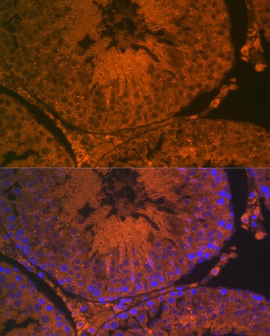
Immunofluorescence analysis of rat testis using ACE2 Rabbit pAb at dilution of 1:100 (40x lens). Blue: DAPI for nuclear staining.

No documents available.