Acetylcholinesterase/ACHE

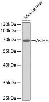
Western blot analysis of extracts of
mouse liver, using ACHE antibody
Secondary antibody: HRP Goat Anti-
Rabbit IgG (H+L) at 1:10000
dilution.
Lysates/proteins: 25ug per lane.
Blocking buffer: 3% nonfat dry milk in
TBST.

Western blot analysis of extracts of mouse liver, using ACHE antibody Secondary antibody: HRP Goat Anti- Rabbit IgG (H+L) at 1:10000 dilution. Lysates/proteins: 25ug per lane. Blocking buffer: 3% nonfat dry milk in TBST.

Catalog Number:AB-84167
Conjugate:Unconjugated
Size:100 ul
Concentration:1mg/ml
Host:Rb
Isotype:IgG
Clone:POLY
Immunogen:A synthetic peptide of human ACHE
Reactivity:Hu, Ms
Applications:Western Blot: 1:200 - 1:2000 Immunohistochemistry: 1:50 - 1:200 Immunofluorescence: 1:50 - 1:200 Flow Cytometry: 1:20 - 1:50
Molecular Weight:68kDa
Purification:Aff. Pur.
Form:Liquid
Synonyms:ACHE;ACEE;ARACHE;N-ACHE;YT

Applications

Western Blot: 1:200 - 1:2000 Immunohistochemistry: 1:50 - 1:200 Immunofluorescence: 1:50 - 1:200 Flow Cytometry: 1:20 - 1:50

Immunogen

A synthetic peptide of human ACHE
Target Background

Acetylcholinesterase hydrolyzes the neurotransmitter, acetylcholine at neuromuscular junctions and brain cholinergic synapses, and thus terminates signal transmission. It is also found on the red blood cell membranes, where it constitutes the Yt blood group antigen. Acetylcholinesterase exists in multiple molecular forms which possess similar catalytic properties, but differ in their oligomeric assembly and mode of cell attachment to the cell surface. It is encoded by the single ACHE gene, and the structural diversity in the gene products arises from alternative mRNA splicing, and post-translational associations of catalytic and structural subunits. The major form of acetylcholinesterase found in brain, muscle and other tissues is the hydrophilic species, which forms disulfide-linked oligomers with collagenous, or lipidcontaining structural subunits. The other, alternatively spliced form, expressed primarily in the erythroid tissues, differs at the C-terminal end, and contains a cleavable hydrophobic peptide with a GPI-anchor site. It associates with the membranes through the phosphoinositide (PI) moieties added post-translationally.

Synonyms:ACHE;ACEE;ARACHE;N-ACHE;YT

Western blot analysis of extracts of mouse liver, using ACHE antibody Secondary antibody: HRP Goat Anti- Rabbit IgG (H+L) at 1:10000 dilution. Lysates/proteins: 25ug per lane. Blocking buffer: 3% nonfat dry milk in TBST.

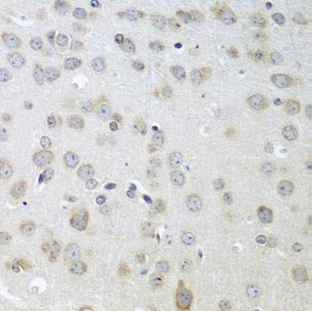
Immunohistochemistry of paraffinembedded mouse brain using ACHE antibody at dilution of 1:100 (40x lens).

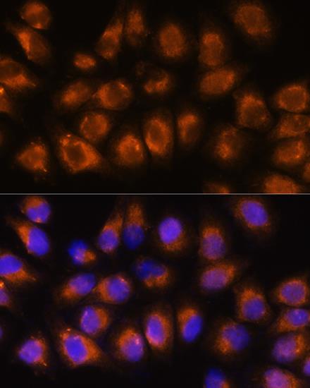
Immunofluorescence analysis of HeLa cells using ACHE Rabbit pAb at dilution of 1:100. Blue: DAPI for nuclear staining.

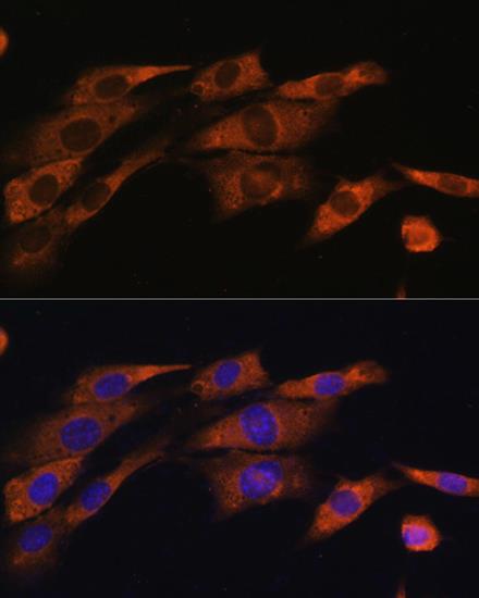
Immunofluorescence analysis of NIH/3T3 cells using ACHE Rabbit pAb at dilution of 1:100. Blue: DAPI for nuclear staining.

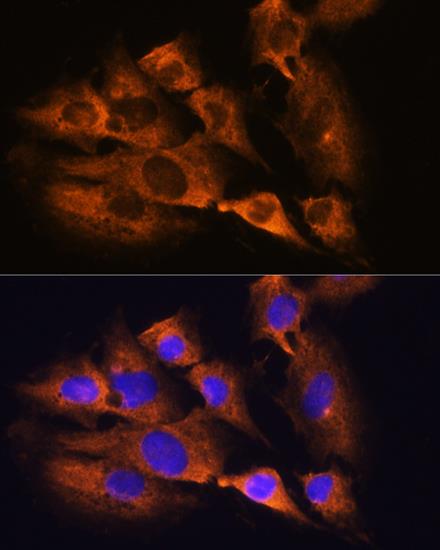
Immunofluorescence analysis of C6 cells using ACHE Rabbit pAb at dilution of 1:100. Blue: DAPI for nuclear staining.

No documents available.