
ACKR3

Western blot analysis of extracts of
various cell lines, using ACKR3
antibody at 1:3000 dilution.
Secondary antibody: HRP
Goat Anti-Rabbit IgG (H+L)
at 1:10000 dilution. Lysates/proteins:
25ug per lane._Blocking buffer: 3%
nonfat dry milk in TBST._Detection:
ECL West Pico Plus.
Exposure time: 90s.

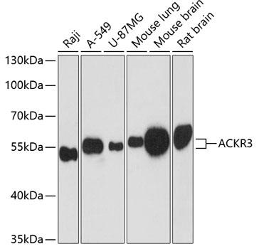
Western blot analysis of extracts of various cell lines, using ACKR3 antibody at 1:3000 dilution. Secondary antibody: HRP Goat Anti-Rabbit IgG (H+L) at 1:10000 dilution. Lysates/proteins: 25ug per lane._Blocking buffer: 3% nonfat dry milk in TBST._Detection: ECL West Pico Plus. Exposure time: 90s.

Catalog Number:AB-84309
Size:100 ug
Concentration:1mg/ml
Host:Rb
Isotype:IgG
Clone:POLY
Immunogen:A synthetic peptide corresponding to a sequence within amino acids 50-150 of human ACKR3
Reactivity:Hu, Ms, Rt
Applications:Western Blot: 1:500 - 1:2000 Immunofluorescence: 1:50 - 1:200
Molecular Weight:55KkDa
Purification:Aff. Pur.
Form:Liquid
Synonyms:ACKR3;CMKOR1;CXC-R7;CXCR-7;CXCR7;GPR159;RDC-1;RDC1

Applications

Western Blot: 1:500 - 1:2000 Immunofluorescence: 1:50 - 1:200

Immunogen

A synthetic peptide corresponding to a sequence within amino acids 50-150 of human ACKR3
Target Background

This gene encodes a member of the G-protein coupled receptor family. Although this protein was earlier thought to be a receptor for vasoactive intestinal peptide (VIP), it is now considered to be an orphan receptor, in that its endogenous ligand has not been identified. The protein is also a coreceptor for human immunodeficiency viruses (HIV). Translocations involving this gene and HMGA2 on chromosome 12 have been observed in lipomas.

Synonyms:ACKR3;CMKOR1;CXC-R7;CXCR-7;CXCR7;GPR159;RDC-1;RDC1

Storage

Store at -20°C, and avoid repeat freeze-thaw cycles.

Buffer

PBS with 0.02% sodium azide,50% glycerol,pH7.3.

Western blot analysis of extracts of various cell lines, using ACKR3 antibody at 1:3000 dilution. Secondary antibody: HRP Goat Anti-Rabbit IgG (H+L) at 1:10000 dilution. Lysates/proteins: 25ug per lane._Blocking buffer: 3% nonfat dry milk in TBST._Detection: ECL West Pico Plus. Exposure time: 90s.

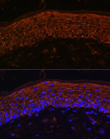
Immunofluorescence analysis of Mouse skin using ACKR3 Rabbit pAb at dilution of 1:100. Blue: DAPI for nuclear staining.

No documents available.