
AKT2 Rabbit Polyclonal Antibody
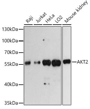
Western blot analysis of extracts of various cell lines, using AKT2 antibody ) at 1:3000 dilution. Secondary antibody: HRP Goat Anti-Rabbit IgG (H+L)at 1:10000 dilution. Lysates/proteins: 25ug per lane. Blocking buffer: 3% nonfat dry milk in TBST. Detection: ECL West Pico Plus. Exposure time: 60s.
| Catalog Number: | AB-83368 |
| Conjugate: | Unconjugated |
| Size: | 100 ug |
| Concentration: | 1mg/ml |
| Host: | Rb |
| Isotype: | IgG |
| Clone: | POLY |
| Immunogen: | A synthetic peptide corresponding to a sequence within amino acids 400 to the Cterminus of human AKT2 |
| Reactivity: | Human, Mouse, Rat, Monkey |
| Applications: | Western Blot: 1:1000 - 1:5000 Immunohistochemistry: 1:50 - 1:100 Immunofluorescence: 1:50 - 1:100 Immunocytochemistry: 1:50 - 1:100 Immunoprecipitation: 1:20 - 1:50 ChIP: 1:20 - 1:100 |
| Molecular Weight: | 60kDa |
| Purification: | Aff. Pur. |
| Form: | Liquid |
| Synonyms: | AKT2;HIHGHH;PKBB;PKBBETA;PRKBB;RAC-BETA |
Applications
Western Blot: 1:1000 - 1:5000 Immunohistochemistry: 1:50 - 1:100 Immunofluorescence: 1:50 - 1:100 Immunocytochemistry: 1:50 - 1:100 Immunoprecipitation: 1:20 - 1:50 ChIP: 1:20 - 1:100
Immunogen
A synthetic peptide corresponding to a sequence within amino acids 400 to the Cterminus of human AKT2