Ampk Alpha1 / PRKAA1

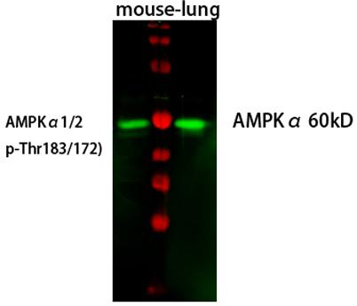
Western Blot analysis of mouse-lung cells using primary
antibody diluted at 1:1000(4°C overnight). Secondary
antibody:Goat Anti-rabbit IgG IRDye 800( diluted at
1:5000, 25°C, 1 hour). Cell lysate was extracted by
Minute™ Plasma Membrane Protein Isolation

Western Blot analysis of mouse-lung cells using primary antibody diluted at 1:1000(4°C overnight). Secondary antibody:Goat Anti-rabbit IgG IRDye 800( diluted at 1:5000, 25°C, 1 hour). Cell lysate was extracted by Minute™ Plasma Membrane Protein Isolation

Catalog Number:AB-84049
Conjugate:Unconjugated
Size:100ug
Concentration:1mg/ml
Host:Rb
Isotype:IgG
Clone:POLY
Immunogen:The antiserum was produced against synthesized peptide derived from human AMPK1. AA range:451-500
Reactivity:Hu, Ms, Rt
Applications:Western Blot: 1/500 - 1/2000 Immunohistochemistry: 1/100 - 1/300 Immunofluorescence: 1/200 - 1/1000 ELISA: 1/10000.
Molecular Weight:65kDa
Purification:The antibody was affinity-purified from rabbit antiserum by affinity-chromatography using epitope-specific immunogen.
Form:Liquid
Synonyms:PRKAA1; AMPK; AMPKa1; protein kinase AMP-activated catalytic subunit alpha 1

Applications

Western Blot: 1/500 - 1/2000 Immunohistochemistry: 1/100 - 1/300 Immunofluorescence: 1/200 - 1/1000 ELISA: 1/10000.

Immunogen

The antiserum was produced against synthesized peptide derived from human AMPK1. AA range:451-500
Target Background

The protein encoded by this gene belongs to the ser/thr protein kinase family. It is the catalytic subunit of the 5'-prime-AMP-activated protein kinase (AMPK). AMPK is a cellular energy sensor conserved in all eukaryotic cells. The kinase activity of AMPK is activated by the stimuli that increase the cellular AMP/ATP ratio. AMPK regulates the activities of a number of key metabolic enzymes through phosphorylation. It protects cells from stresses that cause ATP depletion by switching off ATPconsuming biosynthetic pathways. Alternatively spliced transcript variants encoding distinct isoforms have been observed

Synonyms:PRKAA1; AMPK; AMPKa1; protein kinase AMP-activated catalytic subunit alpha 1

Storage

Store at -20°C. Avoid repeated freeze-thaw cycles.

Buffer

Liquid in PBS containing 50% glycerol, 0.5% BSA and 0.02% sodium azide.

Western Blot analysis of mouse-lung cells using primary antibody diluted at 1:1000(4°C overnight). Secondary antibody:Goat Anti-rabbit IgG IRDye 800( diluted at 1:5000, 25°C, 1 hour). Cell lysate was extracted by Minute™ Plasma Membrane Protein Isolation

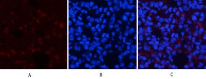
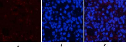
Immunofluorescence analysis of rat-lung tissue. 1,AMPKα1 Polyclonal Antibody(red) was diluted at 1:200(4°C,overnight). 2, Cy3 labled Secondary antibody was diluted at 1:300(room temperature, 50min). 3, Picture B: DAPI(blue) 10min. Picture A:Target.

Immunofluorescence analysis of rat-lung tissue. 1,AMPKα1 Polyclonal Antibody(red) was diluted at 1:200(4°C,overnight). 2, Cy3 labled Secondary antibody was diluted at 1:300(room temperature, 50min).3, Picture B: DAPI(blue) 10min. Picture A:Target. Picture B: DAPI. Picture C: merge of A+B

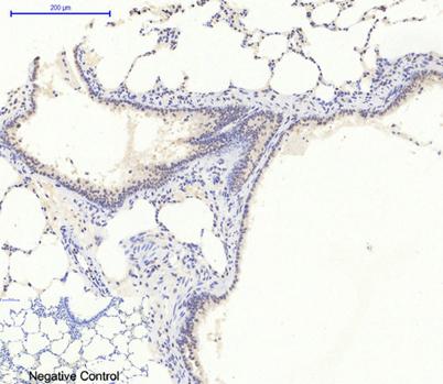
Immunohistochemical analysis of paraffin-embedded Rat-lung tissue. 1,AMPKα1 Polyclonal Antibody was diluted at 1:200(4°C,overnight). 2, Sodium citrate pH 6.0 was used for antibody retrieval(>98°C,20min). 3,Secondary antibody was diluted at 1:200(room tempeRature, 30min). Negative control was used by secondary antibody only.

No documents available.