Anti-GFAP Rabbit Polyclonal Antibody

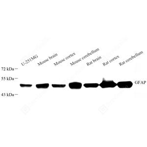
WB analysis of GFAP.Sample: Protein treated by RIPA Lysis Buffer .Blocking buffer: 3% Nonfat dry milk in TBST, RT, 1h.Primary antibody: 1: 1000, 4℃ overnight.Secondary antibody: HRP conjugated Goat Anti-Rabbit lgG , 1: 5000, RT, 1h.

WB analysis of GFAP.Sample: Protein treated by RIPA Lysis Buffer .Blocking buffer: 3% Nonfat dry milk in TBST, RT, 1h.Primary antibody: 1: 1000, 4℃ overnight.Secondary antibody: HRP conjugated Goat Anti-Rabbit lgG , 1: 5000, RT, 1h.

Catalog Number:ABS11096
Conjugate:unconjugated
Size:100 μL
Concentration:1mg/ml
Host:Rabbit
Isotype:IgG
Immunogen:KLH conjugated Synthetic peptide corresponding to Mouse GFAP
Reactivity:Species, Human, Mouse, Rat, Human, Mouse, Rat, Monkey
Applications:Western Blot: 1: 1000-1: 2000 Immunohistochemistry: 1: 500-1: 3000 Immunofluoscence: 1: 500-1: 3000
Purification:Affinity purification
Form:Liquid
Synonyms:GFAP, ALXDRD, glial fibrillary acidic protein, Astrocyte, Intermediate filament protein

Glial fibrillary acidic protein is an intermediate filament (IF) protein that is expressed by numerous cell types of the central nervous system (CNS) including astrocytes and ependymal cells.GFAP has also been found to be expressed in glomeruli and peritubular fibroblasts taken from rat kidneys Leydig cells of the testis in both hamsters and humans, human keratinocytes, human osteocytes and chondrocytes and stellate cells of the pancreas and liver in rats. It is closely related to its non-epithelial family members, vimentin,desmin, and peripherin, which are all involved in the structure and function of the cell’s cytoskeleton.

Applications

Western Blot: 1: 1000-1: 2000 Immunohistochemistry: 1: 500-1: 3000 Immunofluoscence: 1: 500-1: 3000

Immunogen

KLH conjugated Synthetic peptide corresponding to Mouse GFAP
Target Background

Glial fibrillary acidic protein is an intermediate filament (IF) protein that is expressed by numerous cell types of the central nervous system (CNS) including astrocytes and ependymal cells.GFAP has also been found to be expressed in glomeruli and peritubular fibroblasts taken from rat kidneys Leydig cells of the testis in both hamsters and humans, human keratinocytes, human osteocytes and chondrocytes and stellate cells of the pancreas and liver in rats. It is closely related to its non-epithelial family members, vimentin,desmin, and peripherin, which are all involved in the structure and function of the cell’s cytoskeleton.

Synonyms:GFAP, ALXDRD, glial fibrillary acidic protein, Astrocyte, Intermediate filament protein

Storage

Store at -20℃ for one year. Avoid repeated freeze/thaw cycles.

Buffer

PBS with 0.15% ProClin300, 100 μg/mL BSA and 50% glycerol.

WB analysis of GFAP.Sample: Protein treated by RIPA Lysis Buffer .Blocking buffer: 3% Nonfat dry milk in TBST, RT, 1h.Primary antibody: 1: 1000, 4℃ overnight.Secondary antibody: HRP conjugated Goat Anti-Rabbit lgG , 1: 5000, RT, 1h.

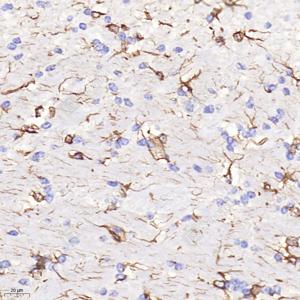
IHC analysis of GFAP.Sample: Rat brain, 4% PFA 12-24h.Antigen retrieval: Citrate buffer , 98℃, 20 min.Primary antibody: 1: 3000, 4℃ overnight.Secondary antibody: HRP conjugated Goat Anti-Rabbit lgG , 1: 200, RT, 1h.

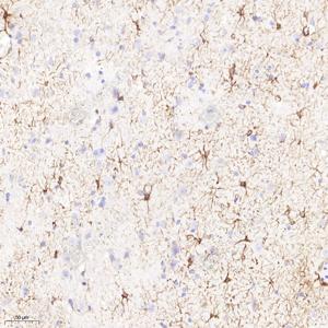
IHC analysis of GFAP.Sample: Mouse brain, 4% PFA 12-24h.Antigen retrieval: Citrate buffer , 98℃, 20 min.Primary antibody: 1: 3000, 4℃ overnight.Secondary antibody: HRP conjugated Goat Anti-Rabbit lgG , 1: 200, RT, 1h.

No documents available.