Anti-ICAM1 Rabbit pAb

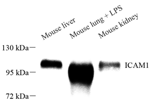
WB analysis of ICAM1 . Sample: Protein treated by RIPA Lysis Buffer . Blocking buffer: 3% Nonfat dry milk in TBST, RT, 1h. Primary antibody: 1: 1000, 4℃ overnight. Secondary antibody: HRP Goat Anti-Rabbit lgG , 1: 3000, RT, 1h.

WB analysis of ICAM1 . Sample: Protein treated by RIPA Lysis Buffer . Blocking buffer: 3% Nonfat dry milk in TBST, RT, 1h. Primary antibody: 1: 1000, 4℃ overnight. Secondary antibody: HRP Goat Anti-Rabbit lgG , 1: 3000, RT, 1h.

Catalog Number:ABS11106
Conjugate:unconjugated
Size:100 μL
Concentration:1mg/ml
Host:Rabbit
Isotype:IgG
Immunogen:KLH conjugated Synthetic peptide corresponding to Mouse ICAM-1
Reactivity:Mouse, Rat
Applications:IHC/IF<br/>1: 500-1: 5000
Molecular Weight:89/100 kDa
Purification:Affinity purification
Form:Liquid

ICAM proteins are ligands for the leukocyte adhesion protein LFA-1 (integrin alpha-L/beta-2). During leukocyte trans-endothelial migration, ICAM1 engagement promotes the assembly of endothelial apical cups through ARHGEF26/SGEF and RHOG activation.(Microbial infection) Acts as a receptor for major receptor group rhinovirus A-B capsid proteins . Acts as a receptor for Coxsackievirus A21 capsid proteins . Upon Kaposis sarcoma-associated herpesvirus/HHV-8 infection, is degraded by viral E3 ubiquitin ligase MIR2, presumably to prevent lysis of infected cells by cytotoxic T-lymphocytes and NK cell . Images

Applications

IHC/IF<br/>1: 500-1: 5000

Immunogen

KLH conjugated Synthetic peptide corresponding to Mouse ICAM-1
Target Background

ICAM proteins are ligands for the leukocyte adhesion protein LFA-1 (integrin alpha-L/beta-2). During leukocyte trans-endothelial migration, ICAM1 engagement promotes the assembly of endothelial apical cups through ARHGEF26/SGEF and RHOG activation.(Microbial infection) Acts as a receptor for major receptor group rhinovirus A-B capsid proteins . Acts as a receptor for Coxsackievirus A21 capsid proteins . Upon Kaposis sarcoma-associated herpesvirus/HHV-8 infection, is degraded by viral E3 ubiquitin ligase MIR2, presumably to prevent lysis of infected cells by cytotoxic T-lymphocytes and NK cell . Images

Storage

Store at -20 ℃ for one year. Avoid repeated freeze/ thaw cycles.

Buffer

PBS with 0.02% sodium azide, 100 μg/ml BSA and 50% glycerol.

WB analysis of ICAM1 . Sample: Protein treated by RIPA Lysis Buffer . Blocking buffer: 3% Nonfat dry milk in TBST, RT, 1h. Primary antibody: 1: 1000, 4℃ overnight. Secondary antibody: HRP Goat Anti-Rabbit lgG , 1: 3000, RT, 1h.

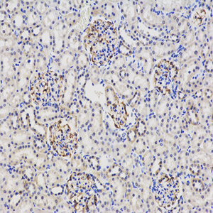
IHC analysis of ICAM1 . Sample: Mouse kidney , 4% PFA 12-24h. Antigen retrieval: Citrate buffer ,98℃,20 min. Blocking buffer: 3% BSA in PBS , RT, 30min. Primary antibody: 1: 6000, 4℃ overnight. Secondary antibody: HRP Goat Anti-Rabbit lgG , 1: 200 RT 1h.

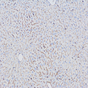
IHC analysis of ICAM1 . Sample: Mouse liver , 4% PFA 12-24h. Antigen retrieval: Citrate buffer ,98℃,20 min. Blocking buffer: 3% BSA in PBS , RT, 30min. Primary antibody: 1: 6000, 4℃ overnight. Secondary antibody: HRP Goat Anti-Rabbit lgG , 1: 200 RT 1h.

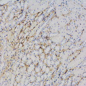
IHC analysis of ICAM1 . Sample: Rat kidney , 4% PFA 12-24h. Antigen retrieval: Citrate buffer ,98℃,20 min. Blocking buffer: 3% BSA in PBS , RT, 30min. Primary antibody: 1: 6000, 4℃ overnight. Secondary antibody: HRP Goat Anti-Rabbit lgG , 1: 200 RT 1h.

No documents available.