AP-1

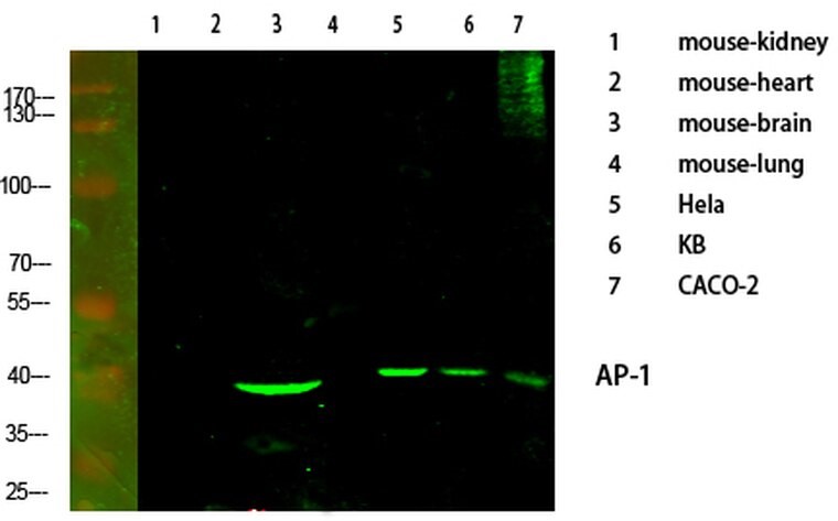
Western blot analysis of various cells using primary antibody diluted at 1:1000 (4°C overnight). Secondary antibody:Goat Anti-rabbit IgG IRDye 800 (diluted at 1:5000, 25°C, 1 hour). Cell lysate was extracted by Minute Plasma Membrane Isolation and Cell Fractionation Kit

Western blot analysis of various cells using primary antibody diluted at 1:1000 (4°C overnight). Secondary antibody:Goat Anti-rabbit IgG IRDye 800 (diluted at 1:5000, 25°C, 1 hour). Cell lysate was extracted by Minute Plasma Membrane Isolation and Cell Fractionation Kit

Catalog Number:AB-84334
Size:100ug
Concentration:1mg/ml
Host:Rb
Isotype:IgG
Clone:POLY
Immunogen:Synthesized peptide derived from human AP-1 around the non-phosphorylation site of T91.
Reactivity:Hu, Ms, Rt
Applications:Western Blot: 1:500-1:2000 Immunofluorescence: 1:50-200 Immunohistochemistry: 1:100-1:300 Immunoprecipitation: 2-5 ug/mg lysate ELISA: 1:20000
Purification:The antibody was affinity-purified from rabbit antiserum by affinity-chromatography using a epitope-specific immunogen.
Form:Liquid

Applications

Western Blot: 1:500-1:2000 Immunofluorescence: 1:50-200 Immunohistochemistry: 1:100-1:300 Immunoprecipitation: 2-5 ug/mg lysate ELISA: 1:20000

Immunogen

Synthesized peptide derived from human AP-1 around the non-phosphorylation site of T91.
Target Background

Storage

Store at -20℃. Avoid freeze / thaw cycles.

Buffer

Liquid in PBS containing 50% glycerol, 0.5% BSA and 0.02% sodium azide.

Western blot analysis of various cells using primary antibody diluted at 1:1000 (4°C overnight). Secondary antibody:Goat Anti-rabbit IgG IRDye 800 (diluted at 1:5000, 25°C, 1 hour). Cell lysate was extracted by Minute Plasma Membrane Isolation and Cell Fractionation Kit

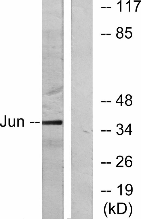
Western blot analysis of lysates from HeLa cells using C-Jun antibody. The lane on the right is blocked with the synthesized peptide.

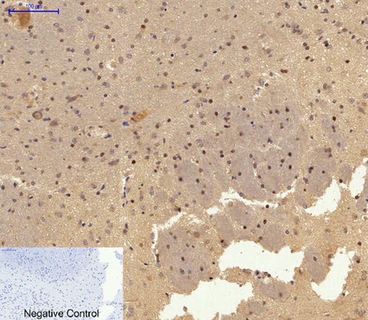
Immunohistochemical analysis of paraffin-embedded Rat-brain tissue. 1, AP-1 Polyclonal Antibody was diluted at 1:200 (4°C, overnight). 2, Sodium citrate pH 6.0 was used for antibody retrieval (>98°C, 20min). 3, Secondary antibody was diluted at 1:200 (room temperature, 30min). Negative control was used by secondary antibody only.

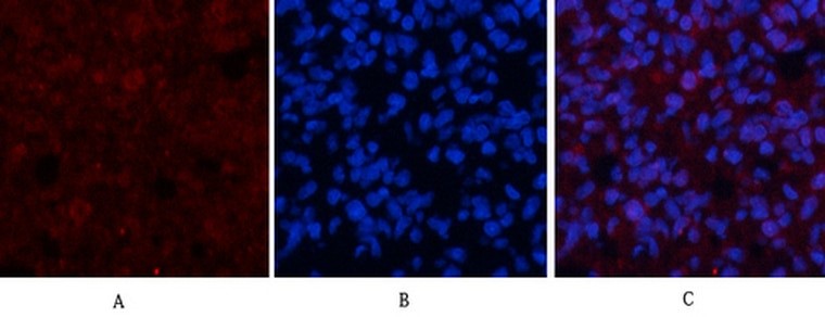
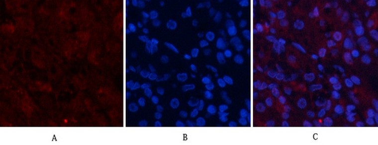
Immunohistochemical analysis of paraffin-embedded Rat-brain tissue. 1, AP-1 Polyclonal Antibody was diluted at 1:200 (4°C, overnight). 2, Sodium citrate pH 6.0 was used for antibody retrieval (>98°C, 20min). 3, Secondary antibody was diluted at 1:200 (room temperature, 30min). Negative control was used by secondary antibody only. Immunofluorescence analysis of human-stomach tissue. 1, AP-1 Polyclonal Antibody (red was diluted at 1:200 (4°C, overnight). 2, Cy3 labled Secondary antibody was diluted at 1:300 (room temperature, 50min).3, Picture B: DAPI (blue) 10min. Picture A:Target. Picture B: DAPI. Picture C: merge of A+B

Immunofluorescence analysis of human-stomach tissue. 1, AP-1 Polyclonal Antibody (red) was diluted at 1:200 (4°C, overnight). 2, Cy3 labled Secondary antibody was diluted at 1:300 (room temperature, 50min).3, Picture B: DAPI (blue) 10min. Picture A:Target. Picture B: DAPI. Picture C: merge of A+B

No documents available.