
APC Anti-Mouse CD279/PD-1 Antibody[29F.1A12]
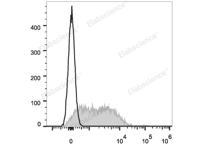
Con-A stimulated C57BL/6 splenocytes (3 days) are stained with APC Anti-Mouse CD279/PD-1 Antibody (filled gray histogram). Unstained splenocytes (empty black histogram) are used as control.
| Catalog Number: | MAB1131UE |
| Conjugate: | APC |
| Size: | 100μg |
| Concentration: | 0.2 mg/mL |
| Host: | Rat |
| Isotype: | Rat IgG2a, κ |
| Clone: | Monoclonal |
| Reactivity: | Mouse |
| Applications: | Each lot of this antibody is quality control tested by flow cytometric analysis. Please check your vial before the experiment. Since applications vary, the appropriate dilutions must be determined for individual use. We suggest each investigator should titrate the reagent to obtain optimal results [The recommended concentration is 0.1-1 μg/10<sup>6</sup> cells in 100 μL volume]. |
| Form: | Liquid |
| Synonyms: | Pd1;Pdcd1 |
CD279, also known as programmed death-1 (PD-1), is a 50-55 kD glycoprotein belonging to the CD28 family of the Ig superfamily. PD-1 is expressed on activated splenic T and B cells and thymocytes. It is induced on activated myeloid cells as well. PD-1 is involved in lymphocyte clonal selection and peripheral tolerance through binding its ligands, B7-H1 (PD-L1) and B7-DC (PD-L2). It has been reported that PD-1 and PD-L1 interactions are critical to positive selection and play a role in shaping the T cell repertoire. PD-L1 negative costimulation is essential for prolonged survival of intratesticular islet allografts.
Applications
Each lot of this antibody is quality control tested by flow cytometric analysis. Please check your vial before the experiment. Since applications vary, the appropriate dilutions must be determined for individual use. We suggest each investigator should titrate the reagent to obtain optimal results [The recommended concentration is 0.1-1 μg/10<sup>6</sup> cells in 100 μL volume].