
ARF6

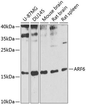
Western blot analysis of extracts of
various cell lines, using ARF6
antibody at 1:1000 dilution.
Secondary antibody: HRP Goat Anti-
Rabbit IgG (H+L) at 1:10000
dilution.
Lysates/proteins: 25ug per lane.
Blocking buffer: 3% nonfat dry milk in
TBST.
Detection: ECL West Pico Plus. 
Exposure time: 10s.

Western blot analysis of extracts of various cell lines, using ARF6 antibody at 1:1000 dilution. Secondary antibody: HRP Goat Anti- Rabbit IgG (H+L) at 1:10000 dilution. Lysates/proteins: 25ug per lane. Blocking buffer: 3% nonfat dry milk in TBST. Detection: ECL West Pico Plus. Exposure time: 10s.

Catalog Number:AB-84267
Size:100ug
Concentration:1mg/ml
Host:Rb
Isotype:IgG
Clone:POLY
Immunogen:Recombinant fusion protein containing a sequence corresponding to amino acids 1-175 of human ARF6
Reactivity:Hu, Ms, Rt
Applications:Western Blot: 1:500 - 1:2000 Immunofluorescence: 1:50 - 1:200 Immunocytochemistry: 1:50-1:200
Molecular Weight:19kDa
Purification:Aff. Pur.
Form:Liquid
Synonyms:ARF6

Applications

Western Blot: 1:500 - 1:2000 Immunofluorescence: 1:50 - 1:200 Immunocytochemistry: 1:50-1:200

Immunogen

Recombinant fusion protein containing a sequence corresponding to amino acids 1-175 of human ARF6
Target Background

This gene encodes a member of the human ARF gene family, which is part of the RAS superfamily. The ARF genes encode small guanine nucleotide-binding proteins that stimulate the ADP-ribosyltransferase activity of cholera toxin and play a role in vesicular trafficking and as activators of phospholipase D. The product of this gene is localized to the plasma membrane, and regulates vesicular trafficking, remodelling of membrane lipids, and signaling pathways that lead to actin remodeling. A pseudogene of this gene is located on chromosome 7.

Synonyms:ARF6

Storage

Store at -20℃. Avoid freeze / thaw cycles.

Buffer

PBS with 0.02% sodium azide,50% glycerol,pH7.3.

Western blot analysis of extracts of various cell lines, using ARF6 antibody at 1:1000 dilution. Secondary antibody: HRP Goat Anti- Rabbit IgG (H+L) at 1:10000 dilution. Lysates/proteins: 25ug per lane. Blocking buffer: 3% nonfat dry milk in TBST. Detection: ECL West Pico Plus. Exposure time: 10s.

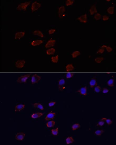
Immunofluorescence analysis of HeLa cells using ARF6 antibody at dilution of 1:100. Blue: DAPI for nuclear staining.

No documents available.