
ARG1

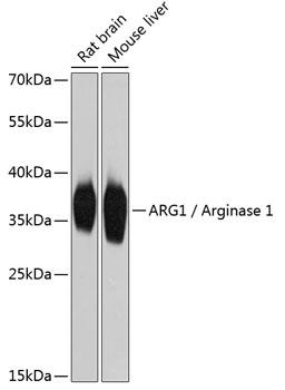
Western blot analysis of extracts of various cell lines, using ARG1 /Arginase 1 Antibody at 1:2000 dilution.
Secondary antibody: HRP Goat Anti-Rabbit IgG (H+L) at 1:10000 dilution.
Lysates/proteins: 25ug per lane.
Blocking buffer: 3% nonfat dry milk in TBST.
Detection: ECL West Pico Plus.    
Exposure time: 1s.

Western blot analysis of extracts of various cell lines, using ARG1 /Arginase 1 Antibody at 1:2000 dilution. Secondary antibody: HRP Goat Anti-Rabbit IgG (H+L) at 1:10000 dilution. Lysates/proteins: 25ug per lane. Blocking buffer: 3% nonfat dry milk in TBST. Detection: ECL West Pico Plus. Exposure time: 1s.

Catalog Number:AB-84248
Size:100 ug
Concentration:1mg/ml
Host:Rb
Isotype:IgG
Clone:POLY
Immunogen:Recombinant fusion protein containing a sequence corresponding to amino acids 1-322 of human ARG1 / Arginase 1
Reactivity:Hu, Ms, Rt
Applications:Western Blot: 1:500 - 1:2000 Immunohistochemistry: 1:50 - 1:200 Immunofluorescence: 1:50 - 1:200
Molecular Weight:36kDa
Purification:Aff. Pur.
Form:Liquid
Synonyms:ARG1;arginase-1

Applications

Western Blot: 1:500 - 1:2000 Immunohistochemistry: 1:50 - 1:200 Immunofluorescence: 1:50 - 1:200

Immunogen

Recombinant fusion protein containing a sequence corresponding to amino acids 1-322 of human ARG1 / Arginase 1

Target Background

Arginase catalyzes the hydrolysis of arginine to ornithine and urea. At least two isoforms of mammalian arginase exist (types I and II) which differ in their tissue distribution, subcellular localization, immunologic crossreactivity and physiologic function. The type I isoform encoded by this gene, is a cytosolic enzyme and expressed predominantly in the liver as a component of the urea cycle. Inherited deficiency of this enzyme results in argininemia, an autosomal recessive disorder characterized by hyperammonemia. Two transcript variants encoding different isoforms have been found for this gene

Synonyms:ARG1;arginase-1

Storage

Store at -20℃. Avoid freeze / thaw cycles.

Buffer

PBS with 0.02% sodium azide,50% glycerol,pH7.3.

Western blot analysis of extracts of various cell lines, using ARG1 /Arginase 1 Antibody at 1:2000 dilution. Secondary antibody: HRP Goat Anti-Rabbit IgG (H+L) at 1:10000 dilution. Lysates/proteins: 25ug per lane. Blocking buffer: 3% nonfat dry milk in TBST. Detection: ECL West Pico Plus. Exposure time: 1s.

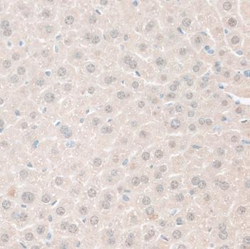
Immunohistochemistry of paraffin embedded mouse liver using ARG1 / Arginase 1 antibody at dilution of 1:100 (40x lens).

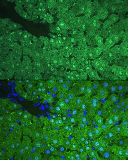
Immunofluorescence analysis of Rat liver using ARG1 / Arginase 1 antibody at dilution of 1:100. Blue: DAPI for nuclear staining.

No documents available.