ATP5A1

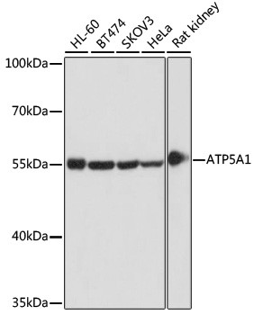
WB analysis of extracts of various cell lines, using ATP5A1 Antibody at 1:1000 dilution.
Secondary antibody: HRP Goat Anti-Rabbit IgG (H+L) at 1:10,000 dilution.
Lysates/proteins: 25ug per lane.
Blocking buffer: 3% nonfat dry milk in TBST.
Detection: ECL West Pico plus Kit .Exposure time: 1s

WB analysis of extracts of various cell lines, using ATP5A1 Antibody at 1:1000 dilution. Secondary antibody: HRP Goat Anti-Rabbit IgG (H+L) at 1:10,000 dilution. Lysates/proteins: 25ug per lane. Blocking buffer: 3% nonfat dry milk in TBST. Detection: ECL West Pico plus Kit .Exposure time: 1s

Catalog Number:MAB-94610
Size:100 ug
Concentration:1mg/ml
Host:Rb
Isotype:IgG
Clone:15H4C4
Immunogen:Recombinant fusion protein containing a sequence corresponding to amino acids 1-280 of human ATP5A1
Reactivity:Hu, Ms, Rt
Applications:Western Blot 1:500 - 1:2000 Immunohistochemistry: 1:50 - 1:200 Immunofluorescence: 1:50 - 1:100 Immunoprecipitation: 1:50 - 1:200
Molecular Weight:53kDa
Purification:Aff. Pur.
Form:Liquid
Synonyms:ATP5A1; ATP5A; ATP5AL2; ATPM; COXPD22; HEL-S-123m; MC5DN4; MOM2; OMR; ORM; hATP1; ATP synthase subunit alpha, mitochondrial

Applications

Western Blot 1:500 - 1:2000 Immunohistochemistry: 1:50 - 1:200 Immunofluorescence: 1:50 - 1:100 Immunoprecipitation: 1:50 - 1:200

Immunogen

Recombinant fusion protein containing a sequence corresponding to amino acids 1-280 of human ATP5A1

Target Background

This gene encodes a subunit of mitochondrial ATP synthase. Mitochondrial ATP synthase catalyzes ATP synthesis, using an electrochemical gradient of protons across the inner membrane during oxidative phosphorylation. ATP synthase is composed of two linked multi-subunit complexes: the soluble catalytic core, F1, and the membrane-spanning component, Fo, comprising the proton channel. The catalytic portion of mitochondrial ATP synthase consists of 5 different subunits (alpha, beta, gamma, delta, and epsilon) assembled with a stoichiometry of 3 alpha, 3 beta, and a single representative of the other 3. The proton channel consists of three main subunits (a, b, c). This gene encodes the alpha subunit of the catalytic core. Alternatively spliced transcript variants encoding the different isoforms have been identified. Pseudogenes of this gene are located on chromosomes 9, 2, and 16.

Synonyms:ATP5A1; ATP5A; ATP5AL2; ATPM; COXPD22; HEL-S-123m; MC5DN4; MOM2; OMR; ORM; hATP1; ATP synthase subunit alpha, mitochondrial

Storage

Store at -20℃. Avoid freeze / thaw cycles.

Buffer

PBS with 0.02% sodium azide, 50% glycerol, pH7.

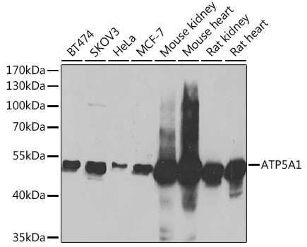
WB analysis of extracts of various cell lines, using ATP5A1 Antibody at 1:1000 dilution. Secondary antibody: HRP Goat Anti-Rabbit IgG (H+L) at 1:10,000 dilution. Lysates/proteins: 25ug per lane. Blocking buffer: 3% nonfat dry milk in TBST. Detection: ECL West Pico plus Kit .Exposure time: 1s

WB analysis of extracts of various cells, using ATP5A1 antibody at 1:1000 dilution ratio through one-step method. Secondary antibody: HRP Goat Anti-Rabbit IgG (H+L) at 1:10000 dilution. Lysates/proteins: 25ug per lane. Blocking buffer: 3% nonfat dry milk in TBST. Detection: ECL West Pico Kit Exposure time: 1s

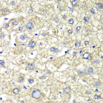
immunohistochemistry of paraffin-embedded human liver injury using ATP5A1 antibody at dilution of 1:100

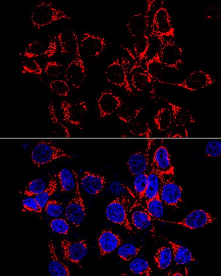
Immunofluorescence analysis of HeLa cells using ATP5A1 antibody at dilution of 1:100 (60x lens). Blue: DAPI for nuclear staining.

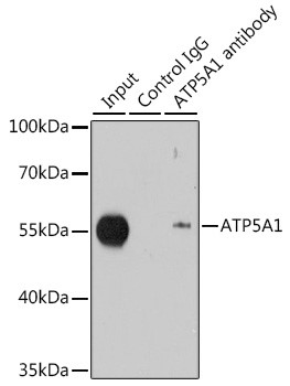
Immunoprecipitation analysis of 200ug extracts of MCF-7 cells using 1ug ATP5A1 antibody .WB was performed from the immunoprecipitate using ATP5A1 antibody at a dilition of 1:1000.

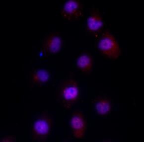
Review for ATP5A1 Rabbit mAb Experiment Type: Immunofluorescence(IF) Sample: HK2 Description: Immunofluorescence analysis of HK2 cells using ATP5A1 antibody at 1:100 dilution. The positive signal is Golgi staining.

No documents available.